Gurel Zafer, Sieg Kelsey M, Shallow Keegan D, Sorenson Christine M, Sheibani Nader
Department of Ophthalmology and Visual Sciences, University of Wisconsin School of Medicine and Public Health, Madison, WI 53792-4673, USA.
Mol Vis. 2013 May 21;19:1047-59. Print 2013.
Hyperglycemia activates several metabolic pathways, including the hexosamine biosynthetic pathway. Uridine diphosphate N-acetylglucosamine (GlcNAc) is the product of the hexosamine biosynthetic pathway and the substrate for O-linked GlcNAc (O-GlcNAc) modification. This modification affects a wide range of proteins by altering their activity, cellular localization, and/or protein interactions. However, the role O-GlcNAcylation may play in normal postnatal retinal vascular development and in the ocular complications of diabetes, including diabetic retinopathy, requires further investigation.
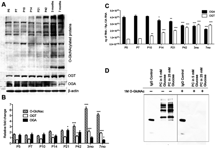
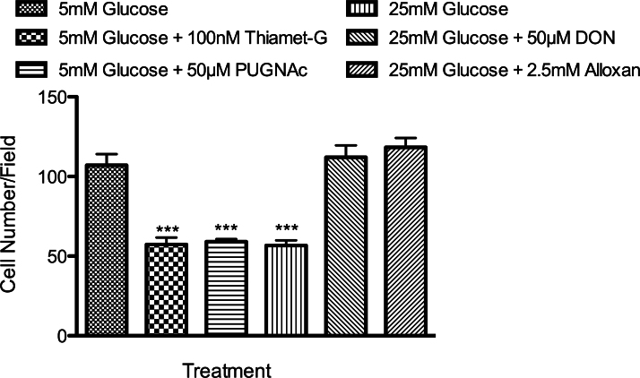
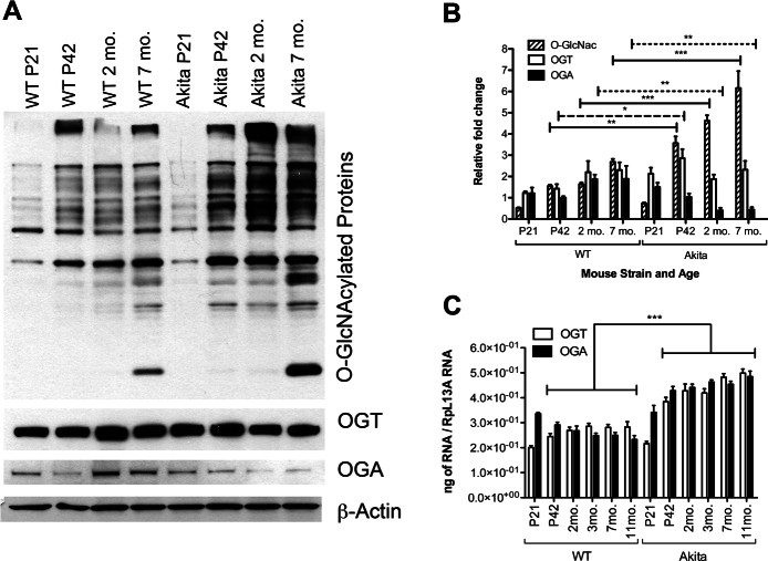
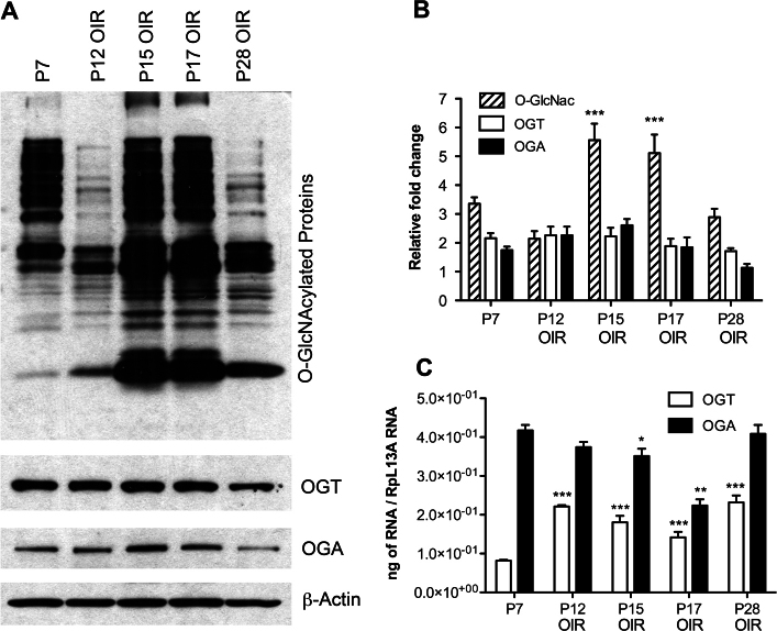
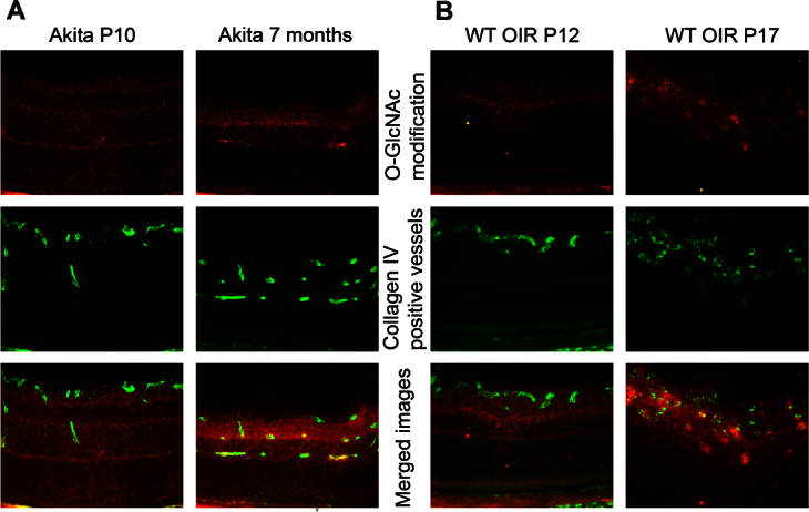
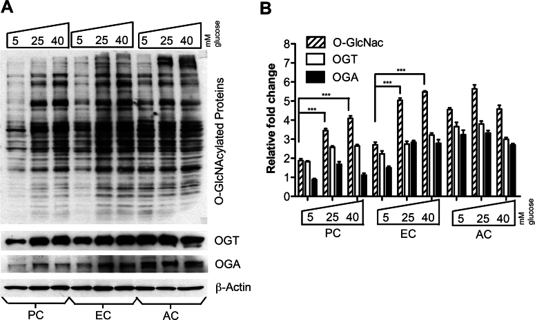
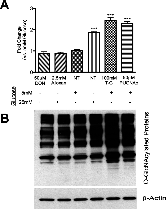
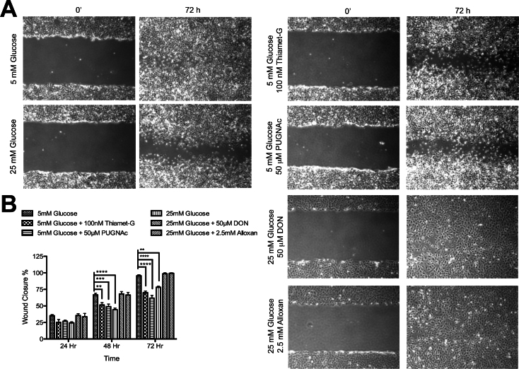
The total levels of O-GlcNAc-modified proteins were evaluated by western blot analysis of lysates prepared from retinas obtained at different days during postnatal retinal vascularization and oxygen-induced ischemic retinopathy. Similar experiments were performed with retinal lysate prepared from diabetic Ins2(Akita/+) mice with different durations of diabetes and retinal vascular cells cultured under various glucose conditions. The localization of O-GlcNAc-modified proteins in the retinal vasculature was confirmed by immunofluorescence staining. The impact of altered O-GlcNAcylation on the migration of retinal vascular cells was determined using scratch wound and transwell migration assays.
We detected an increase in protein O-GlcNAcylation during mouse postnatal retinal vascularization and aging, in part through the regulation of the enzymes that control this modification. The study of the diabetic Ins2(Akita/+) mouse retina showed an increase in the O-GlcNAc modification of retinal proteins. We also observed an increase in retinal O-GlcNAcylated protein levels during the neovascularization phase of oxygen-induced ischemic retinopathy. Our fluorescence microscopy data confirmed that the alterations in retinal O-GlcNAcylation are similarly represented in the retinal vasculature and in retinal pericytes and endothelial cells. Particularly, the migration of retinal pericytes, but not retinal endothelial cells, was attenuated by increased O-GlcNAc modification.
The O-GlcNAc modification pattern changes during postnatal retinal vascular development and neovascularization, and its dysregulation under hyperglycemia and/or ischemia may contribute to the pathogenesis of the diabetic retinopathy and retinal neovascularization.
高血糖会激活多种代谢途径,包括己糖胺生物合成途径。尿苷二磷酸N - 乙酰葡糖胺(GlcNAc)是己糖胺生物合成途径的产物,也是O - 连接的N - 乙酰葡糖胺(O - GlcNAc)修饰的底物。这种修饰通过改变蛋白质的活性、细胞定位和/或蛋白质相互作用来影响多种蛋白质。然而,O - GlcNAc糖基化在正常出生后视网膜血管发育以及糖尿病眼部并发症(包括糖尿病视网膜病变)中可能发挥的作用,仍需进一步研究。
通过对出生后视网膜血管化不同阶段以及氧诱导缺血性视网膜病变时获取的视网膜裂解物进行蛋白质印迹分析,评估O - GlcNAc修饰蛋白的总水平。对不同糖尿病病程的糖尿病Ins2(Akita / +)小鼠的视网膜裂解物以及在不同葡萄糖条件下培养的视网膜血管细胞进行了类似实验。通过免疫荧光染色确认O - GlcNAc修饰蛋白在视网膜血管系统中的定位。使用划痕伤口实验和Transwell迁移实验确定O - GlcNAc糖基化改变对视网膜血管细胞迁移的影响。
我们检测到在小鼠出生后视网膜血管化和衰老过程中,蛋白质O - GlcNAc糖基化增加,部分原因是通过调控控制这种修饰的酶。对糖尿病Ins2(Akita / +)小鼠视网膜的研究显示,视网膜蛋白的O - GlcNAc修饰增加。我们还观察到在氧诱导缺血性视网膜病变的新生血管形成阶段,视网膜O - GlcNAc化蛋白水平增加。我们的荧光显微镜数据证实,视网膜O - GlcNAc糖基化的改变在视网膜血管系统以及视网膜周细胞和内皮细胞中表现相似。特别是,O - GlcNAc修饰增加会减弱视网膜周细胞的迁移,但不会影响视网膜内皮细胞的迁移。
出生后视网膜血管发育和新生血管形成过程中,O - GlcNAc修饰模式会发生变化,在高血糖和/或缺血情况下其失调可能导致糖尿病视网膜病变和视网膜新生血管形成的发病机制。