Department of Endocrinology, Jinshan Hospital, Fudan University, 1508 Longhang Road, Shanghai 201508, PR China.
J Neuroinflammation. 2013 Jun 4;10:69. doi: 10.1186/1742-2094-10-69.
Tumor necrosis factor-α (TNF-α) is an important inflammatory factor produced by activated macrophages and monocytes and plays an important role in the pathogenesis of diabetic peripheral neuropathy (DPN). To evaluate the effect of TNF-α signaling suppression and the potential of TNF-α in the treatment of DPN, a recombinant human TNF-α receptor-antibody fusion protein (rhTNFR:Fc) was used. We focused on the pathophysiology of the sciatic nerve and examined the expression of myelin basic protein (MBP) under DPN status with or without TNF-α inhibition.
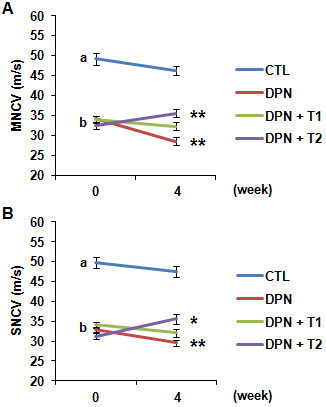
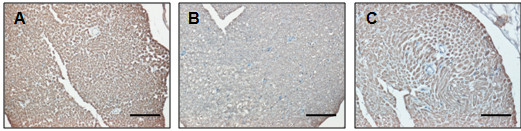
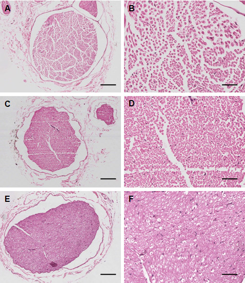
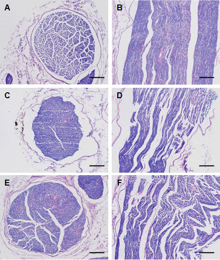
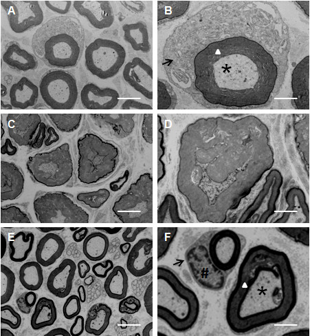
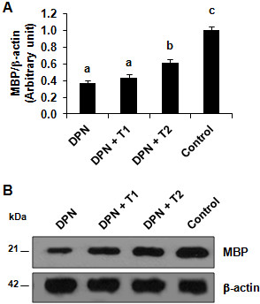
The DPN rat model was generated by intraperitoneal injection of streptozotocin and by feeding with a high-fat, high-sugar diet. The nerve conduction velocity (NCV) in sciatic nerve of rat was monitored over a period of four weeks. The histopathological changes in nerve tissue were examined through traditional tissue histology and ultrastructure transmission electron microscopy (TEM). The expression of MBP was examined through western blot analysis.
The DPN induced rats showed significant signs of nerve damage including lower NCV, demyelination of nerve fibers, disorganization of lamellar and axonal structures, and decreased expression of MBP in the nerve tissue. The inhibition of TNF-α in the DPN rats resulted in a significant recovery from those symptoms compared to the DPN rats.
Our study demonstrates that TNF-α plays a key role in the pathogenesis of DPN and its inhibition by rhTNFR:Fc can prove to be a useful therapeutic strategy for the treatment of and/or prevention from DPN symptoms.
肿瘤坏死因子-α(TNF-α)是一种由激活的巨噬细胞和单核细胞产生的重要炎症因子,在糖尿病周围神经病变(DPN)的发病机制中发挥重要作用。为了评估 TNF-α信号抑制的效果以及 TNF-α在治疗 DPN 中的潜力,使用了一种重组人 TNF-α受体-抗体融合蛋白(rhTNFR:Fc)。我们专注于坐骨神经的病理生理学,研究了 DPN 状态下以及 TNF-α抑制情况下髓鞘碱性蛋白(MBP)的表达。
通过腹腔注射链脲佐菌素并给予高脂肪、高糖饮食,建立 DPN 大鼠模型。监测大鼠坐骨神经的神经传导速度(NCV)在四周内的变化。通过传统组织学和超微结构透射电子显微镜(TEM)检查神经组织的病理变化。通过 Western blot 分析检测 MBP 的表达。
DPN 诱导的大鼠表现出明显的神经损伤迹象,包括 NCV 降低、神经纤维脱髓鞘、板层和轴突结构紊乱以及神经组织中 MBP 表达减少。与 DPN 大鼠相比,DPN 大鼠中 TNF-α 的抑制导致这些症状得到了显著恢复。
我们的研究表明 TNF-α在 DPN 的发病机制中起关键作用,rhTNFR:Fc 的抑制可能成为治疗和/或预防 DPN 症状的有效治疗策略。