Laboratory of Molecular Neurobiology and Biophysics, The Rockefeller University, 1230 York Avenue, New York, New York 10065, USA.
Nature. 2013 Jun 13;498(7453):190-7. doi: 10.1038/nature12241. Epub 2013 Jun 5.
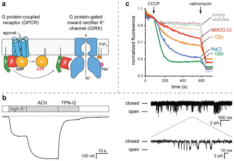
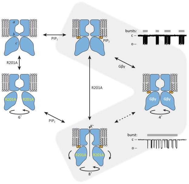
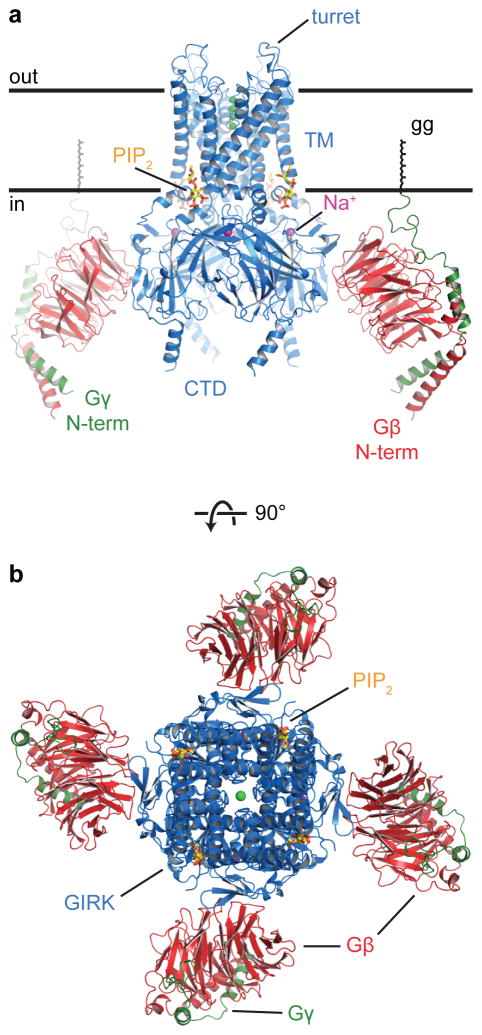
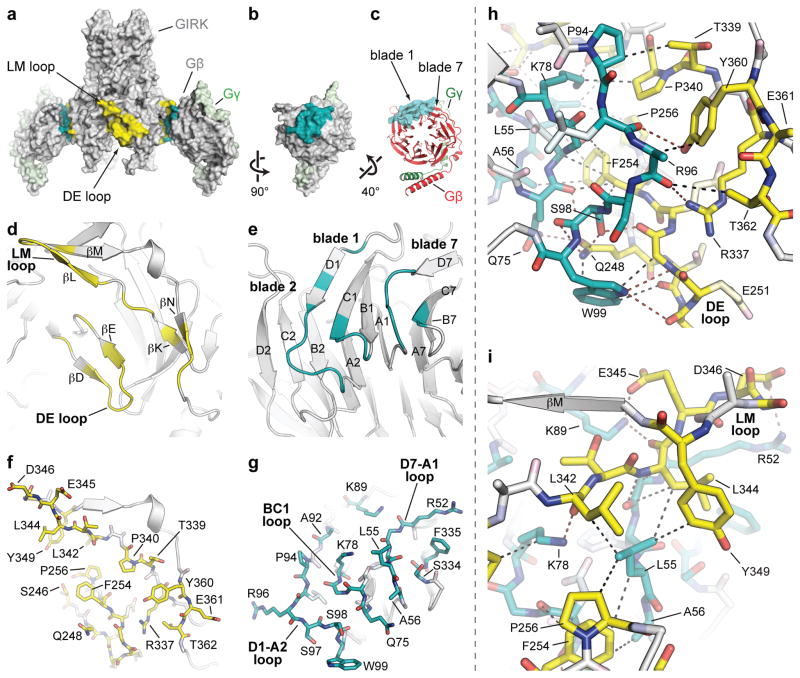
G-protein-gated inward rectifier K(+) (GIRK) channels allow neurotransmitters, through G-protein-coupled receptor stimulation, to control cellular electrical excitability. In cardiac and neuronal cells this control regulates heart rate and neural circuit activity, respectively. Here we present the 3.5 Å resolution crystal structure of the mammalian GIRK2 channel in complex with βγ G-protein subunits, the central signalling complex that links G-protein-coupled receptor stimulation to K(+) channel activity. Short-range atomic and long-range electrostatic interactions stabilize four βγ G-protein subunits at the interfaces between four K(+) channel subunits, inducing a pre-open state of the channel. The pre-open state exhibits a conformation that is intermediate between the closed conformation and the open conformation of the constitutively active mutant. The resultant structural picture is compatible with 'membrane delimited' activation of GIRK channels by G proteins and the characteristic burst kinetics of channel gating. The structures also permit a conceptual understanding of how the signalling lipid phosphatidylinositol-4,5-bisphosphate (PIP2) and intracellular Na(+) ions participate in multi-ligand regulation of GIRK channels.
G 蛋白门控内向整流钾 (GIRK) 通道允许神经递质通过 G 蛋白偶联受体的刺激来控制细胞的电兴奋性。在心脏和神经元细胞中,这种控制分别调节心率和神经回路活动。在这里,我们展示了哺乳动物 GIRK2 通道与 βγ G 蛋白亚基复合物的 3.5Å 分辨率晶体结构,这是连接 G 蛋白偶联受体刺激与 K(+) 通道活性的中央信号复合物。短程原子和远程静电相互作用将四个βγ G 蛋白亚基稳定在四个 K(+) 通道亚基的界面之间,诱导通道的预开放状态。预开放状态表现出一种构象,介于关闭构象和组成型激活突变体的开放构象之间。由此产生的结构图像与 G 蛋白通过“膜限制”激活 GIRK 通道以及通道门控的特征爆发动力学兼容。这些结构还允许我们从概念上理解信号脂质磷脂酰肌醇-4,5-二磷酸 (PIP2) 和细胞内 Na(+) 离子如何参与 GIRK 通道的多配体调节。