Department of Clinic and Surgery, Area Stomatology, Alfenas Federal University, Alfenas, Minas Gerais, Brazil.
Head Face Med. 2013 Jun 11;9:15. doi: 10.1186/1746-160X-9-15.
Peripheral odontoma arising in the extraosseous soft tissues is rare and if not removed early, may enlarge over time and eventually erupt in the oral cavity.
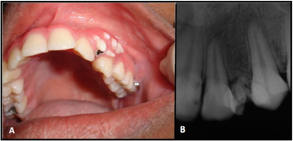
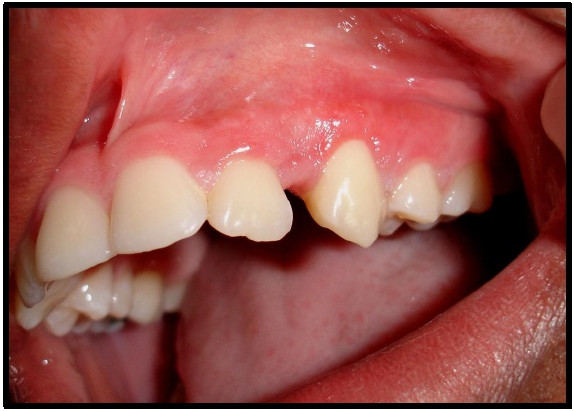
A 15-year-old girl presented with "denticles on the gingiva". During the intraoral examination, seven small tooth-like structures were found. These were exposed in the anterior left gingiva between the permanent maxillary lateral incisor and canine teeth, and the left first premolar was absent. Radiographic examination revealed irregular tooth-like structures without evidence of bone involvement.
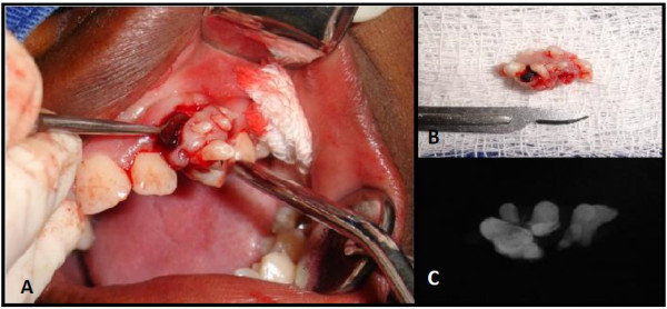
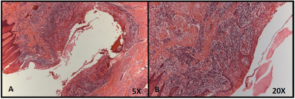
The lesion was surgically removed, and the specimens were analyzed histopathologically. The diagnosis of compound odontoma was established.
This is the twelfth reported case of peripheral odontoma in the gingiva and the first one that erupted in the oral cavity.
发生在骨外软组织中的外周性牙瘤非常罕见,如果不早期切除,可能会随时间逐渐增大,最终在口腔内萌出。
一名 15 岁女孩因“牙龈上有小牙”就诊。口腔检查发现 7 个类似牙的小结构。这些结构在前部左侧牙龈中暴露,位于上颌恒侧切牙和尖牙之间,且左侧第一前磨牙缺失。影像学检查显示不规则的类似牙的结构,无骨受累的证据。
病变被手术切除,标本进行了组织病理学分析。诊断为混合性牙瘤。
这是第十二例报告的发生在牙龈的外周性牙瘤病例,也是首例萌出于口腔的病例。