Department of Ophthalmology, Medical College of Wisconsin, Milwaukee, Wisconsin.
JAMA Ophthalmol. 2013 Sep;131(9):1207-15. doi: 10.1001/jamaophthalmol.2013.387.
Demonstrating the utility of adaptive optics scanning light ophthalmoscopy (AOSLO) to assess outer retinal structure in Best vitelliform macular dystrophy (BVMD).
To characterize outer retinal structure in BVMD using spectral-domain optical coherence tomography (SD-OCT) and AOSLO.
DESIGN, SETTING, AND PARTICIPANTS: Prospective, observational case series. Four symptomatic members of a family with BVMD with known BEST1 mutation were recruited at the Advanced Ocular Imaging Program research lab at the Medical College of Wisconsin Eye Institute, Milwaukee.
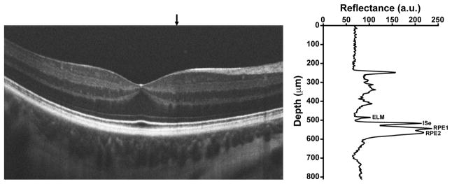
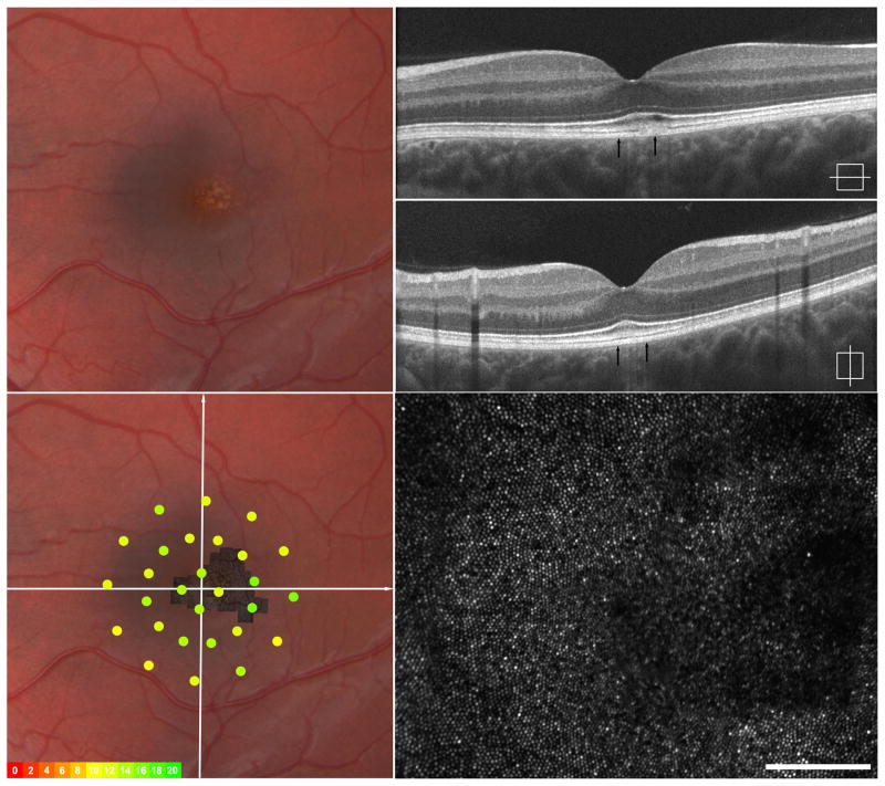
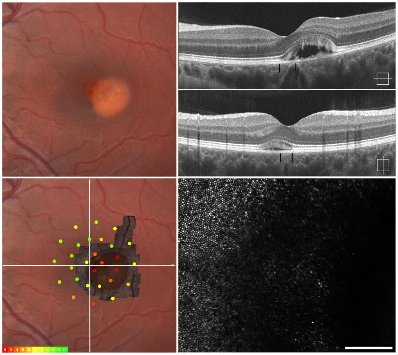
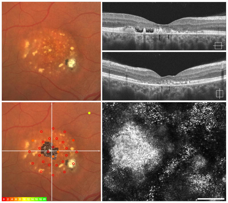
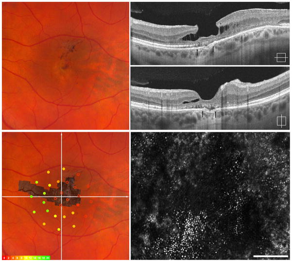
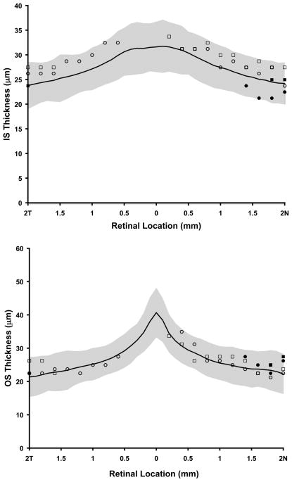
Thickness of 2 outer retinal layers corresponding to photoreceptor inner and outer segments was measured using SD-OCT. Photoreceptor mosaic AOSLO images within and around visible lesions were obtained, and cone density was assessed in 2 subjects.
Photoreceptor structure.
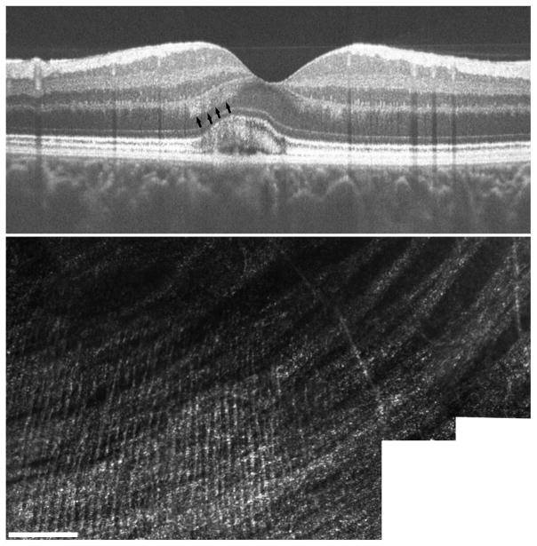
Each subject was at a different stage of BVMD, with photoreceptor disruption evident by AOSLO at all stages. When comparing SD-OCT and AOSLO images from the same location, AOSLO images allowed for direct assessment of photoreceptor structure. A variable degree of retained photoreceptors was seen within all lesions. The photoreceptor mosaic immediately adjacent to visible lesions appeared contiguous and was of normal density. Fine hyperreflective structures were visualized by AOSLO, and their anatomical orientation and size were consistent with Henle fibers.
AND RELEVANCE: The AOSLO findings indicate that substantial photoreceptor structure persists within active lesions, accounting for good visual acuity in these patients. Despite previous reports of diffuse photoreceptor outer segment abnormalities in BVMD, our data reveal normal photoreceptor structure in areas adjacent to clinical lesions. This study demonstrates the utility of AOSLO for understanding the spectrum of cellular changes that occur in inherited degenerations such as BVMD. Photoreceptors are often significantly affected at various stages of inherited degenerations, and these changes may not be readily apparent with current clinical imaging instrumentation.
展示自适应光学扫描检眼镜(AOSLO)在评估 Best 卵黄样黄斑营养不良(BVMD)的外视网膜结构中的效用。
使用谱域光相干断层扫描(SD-OCT)和 AOSLO 来描绘 BVMD 的外视网膜结构。
设计、设置和参与者:前瞻性观察性病例系列。威斯康星医学院眼科研究所高级眼部成像计划研究实验室招募了 4 名有已知 BEST1 突变的 BVMD 症状家族成员。
使用 SD-OCT 测量与光感受器内、外节相对应的 2 个外视网膜层的厚度。在可见病变内和周围获得光感受器镶嵌 AOSLO 图像,并在 2 名受试者中评估锥体细胞密度。
光感受器结构。
每个受试者处于 BVMD 的不同阶段,AOSLO 在所有阶段均显示光感受器破坏。当比较同一位置的 SD-OCT 和 AOSLO 图像时,AOSLO 图像允许直接评估光感受器结构。在所有病变中都可见到一定程度的保留光感受器。紧邻可见病变的光感受器镶嵌物似乎连续,且密度正常。AOSLO 可观察到细微的高反射结构,其解剖方向和大小与 Henle 纤维一致。
和相关性:AOSLO 的发现表明,在活跃病变内存在大量的光感受器结构,这可以解释这些患者良好的视力。尽管之前有报道称 BVMD 存在弥漫性光感受器外节异常,但我们的数据显示,在临床病变周围的区域,光感受器结构正常。本研究证明了 AOSLO 用于理解在遗传性变性(如 BVMD)中发生的细胞变化谱的效用。在遗传性变性的各个阶段,光感受器通常受到严重影响,而这些变化可能不易通过当前的临床成像仪器察觉。