Stem Bank Company, Busan, Korea.
Department of Molecular Biology and Immunology, College of Medicine, Kosin University, Busan, Korea.
Sci Rep. 2020 Jul 24;10(1):12448. doi: 10.1038/s41598-020-69020-z.
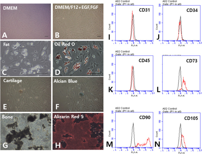
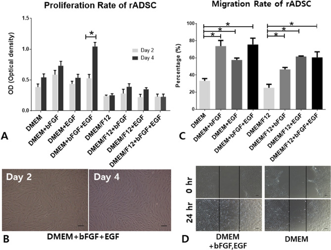
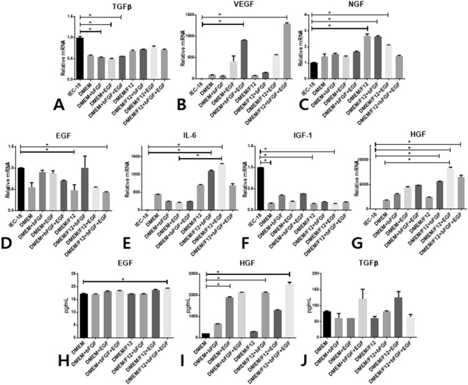
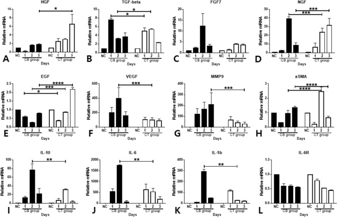
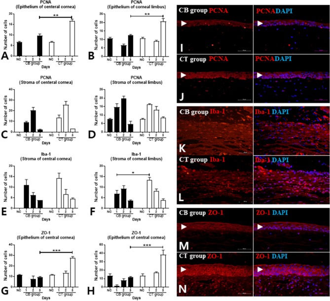
Corneal chemical burns can lead to blindness following serious complications. As most of these complications are caused by failure of reepithelization during the acute phase, treatment at this stage is critical. Although there have been some studies on corneal injury recovery using adipose tissue-derived stem cells (ADSCs), none has reported the effect of topical cell-free conditioned culture media (CM) derived from ADSCs on corneal epithelial regeneration. Here, the best conditions for CM were selected and used for in vitro and in vivo experiments. Corneal burn in rats was induced using 100% alcohol. The chosen CM was administered to corneal burn rats (CM-treated [CT] group) four times a day for three days and this group was compared with the normal control and corneal burn (CB) groups. Biomicroscopic fluorescence images and the actual physical corneas were taken over time and used for analysis. mRNA levels of hepatocyte growth factor and epidermal growth factor (EGF) were significantly increased, whereas those of vascular endothelial growth factor, interleukin (IL)-1β, IL-6, IL-10, and matrix metalloproteinase-9 were significantly decreased in the CT group compared with those in the CB group. The numbers of proliferating cell nuclear antigen- and zonular occludens-1-positive cells in the CT group were significantly higher than those in the CB group. The macrophage-infiltrating corneas in the CT group expressed significantly more of the M2 marker arginase than corneas in the CB group. Optimal CM (× 0.5 concentration) treatment significantly accelerated the migration of corneal epithelial cells and induced upregulation of the expression of IL-6, EGF, and C-X-C chemokine receptor type 4 mRNAs. Overall, in this study, topical administration of cell-free CM promoted regeneration of the corneal epithelium after induction of chemical burns.
角膜化学烧伤可导致严重并发症后失明。由于这些并发症大多是由于急性阶段的再上皮化失败引起的,因此该阶段的治疗至关重要。尽管已经有一些关于脂肪组织来源的干细胞(ADSCs)用于角膜损伤恢复的研究,但没有报道过源自 ADSC 的无细胞条件培养基(CM)对角膜上皮再生的影响。在这里,选择了最佳的 CM 条件,并将其用于体外和体内实验。使用 100%酒精诱导大鼠角膜烧伤。将选择的 CM 每天四次施用于角膜烧伤大鼠(CM 处理[CT]组),持续三天,并将该组与正常对照组和角膜烧伤(CB)组进行比较。随着时间的推移,进行生物显微镜荧光图像和实际物理角膜的拍摄和分析。与 CB 组相比,CT 组的肝细胞生长因子和表皮生长因子(EGF)的 mRNA 水平显著增加,而血管内皮生长因子、白细胞介素(IL)-1β、IL-6、IL-10 和基质金属蛋白酶-9 的 mRNA 水平显著降低。CT 组的增殖细胞核抗原和紧密连接蛋白-1 阳性细胞数量明显高于 CB 组。CT 组的巨噬细胞浸润角膜表达的 M2 标志物精氨酸酶明显多于 CB 组。最佳 CM(×0.5 浓度)处理显著加速了角膜上皮细胞的迁移,并诱导了 IL-6、EGF 和 C-X-C 趋化因子受体 4 mRNAs 的表达上调。总的来说,在这项研究中,局部给予无细胞 CM 可促进化学烧伤后角膜上皮的再生。