• 文献检索
  • 文档翻译
  • 深度研究
  • 学术资讯
  • Suppr Zotero 插件Zotero 插件
  • 邀请有礼
  • 套餐&价格
  • 历史记录
应用&插件
Suppr Zotero 插件Zotero 插件浏览器插件Mac 客户端Windows 客户端微信小程序
定价
高级版会员购买积分包购买API积分包
服务
文献检索文档翻译深度研究API 文档MCP 服务
关于我们
关于 Suppr公司介绍联系我们用户协议隐私条款
关注我们

Suppr 超能文献

核心技术专利:CN118964589B侵权必究
粤ICP备2023148730 号-1Suppr @ 2026

文献检索

告别复杂PubMed语法,用中文像聊天一样搜索,搜遍4000万医学文献。AI智能推荐,让科研检索更轻松。

立即免费搜索

文件翻译

保留排版,准确专业,支持PDF/Word/PPT等文件格式,支持 12+语言互译。

免费翻译文档

深度研究

AI帮你快速写综述,25分钟生成高质量综述,智能提取关键信息,辅助科研写作。

立即免费体验

格雷研究所开放高内涵荧光寿命显微镜。

The Gray Institute 'open' high-content, fluorescence lifetime microscopes.

机构信息

Gray Institute for Radiation Oncology & Biology, Dept. Of Oncology, University of Oxford, Oxford, UK.

出版信息

J Microsc. 2013 Aug;251(2):154-67. doi: 10.1111/jmi.12057. Epub 2013 Jun 12.

DOI:10.1111/jmi.12057
PMID:23772985
原文链接:https://pmc.ncbi.nlm.nih.gov/articles/PMC3910159/
Abstract

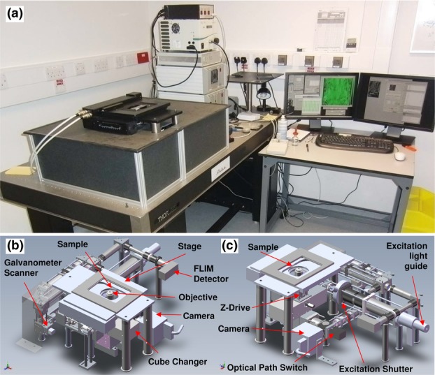
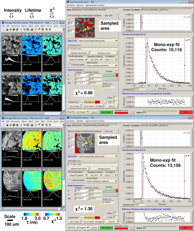
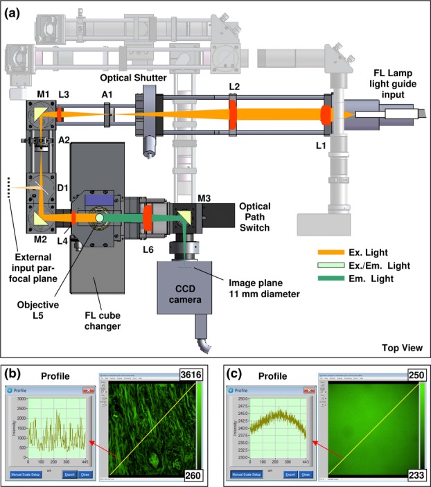
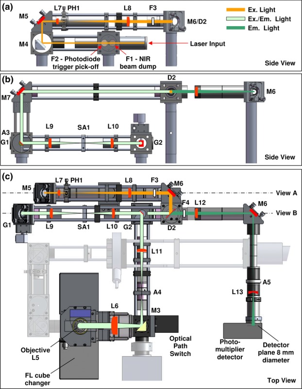
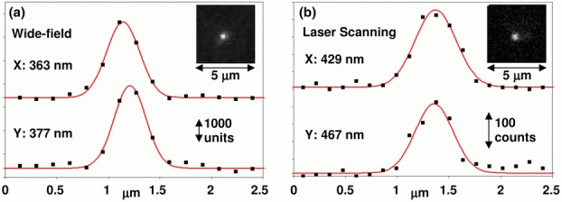
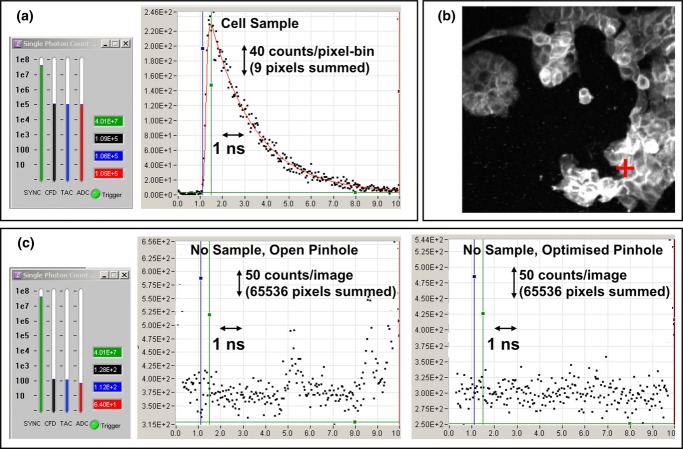
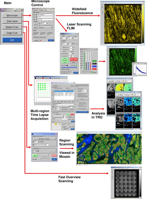
We describe a microscopy design methodology and details of microscopes built to this 'open' design approach. These demonstrate the first implementation of time-domain fluorescence microscopy in a flexible automated platform with the ability to ease the transition of this and other advanced microscopy techniques from development to use in routine biology applications. This approach allows easy expansion and modification of the platform capabilities, as it moves away from the use of a commercial, monolithic, microscope body to small, commercial off-the-shelf and custom made modular components. Drawings and diagrams of our microscopes have been made available under an open license for noncommercial use at http://users.ox.ac.uk/~atdgroup. Several automated high-content fluorescence microscope implementations have been constructed with this design framework and optimized for specific applications with multiwell plates and tissue microarrays. In particular, three platforms incorporate time-domain FLIM via time-correlated single photon counting in an automated fashion. We also present data from experiments performed on these platforms highlighting their automated wide-field and laser scanning capabilities designed for high-content microscopy. Devices using these designs also form radiation-beam 'end-stations' at Oxford and Surrey Universities, showing the versatility and extendibility of this approach.

摘要

我们描述了一种显微镜设计方法和根据该“开放式”设计方法制造的显微镜的详细信息。这些显微镜展示了时域荧光显微镜在灵活自动化平台中的首次实现,该平台具有将这种技术和其他先进显微镜技术从开发阶段轻松过渡到常规生物学应用的能力。这种方法允许轻松扩展和修改平台功能,因为它不再使用商业的、单片的显微镜主体,而是使用小型的、商用现货的和定制的模块化组件。我们的显微镜的图纸和图表已根据开放许可证提供,可供非商业用途在 http://users.ox.ac.uk/~atdgroup 上使用。已经使用该设计框架构建了几个自动化高通量荧光显微镜实现,并针对多孔板和组织微阵列进行了优化。特别是,三个平台以自动化方式通过时间相关单光子计数实现了时域 FLIM。我们还展示了在这些平台上进行的实验数据,突出了它们为高通量显微镜设计的自动化宽场和激光扫描功能。在牛津大学和萨里大学,使用这些设计的设备也形成了辐射束“终端站”,展示了这种方法的多功能性和可扩展性。

https://cdn.ncbi.nlm.nih.gov/pmc/blobs/348d/3910159/8ea09a7c1ad4/jmi0251-0154-f8.jpg
https://cdn.ncbi.nlm.nih.gov/pmc/blobs/348d/3910159/6af2a0e044ae/jmi0251-0154-f1.jpg
https://cdn.ncbi.nlm.nih.gov/pmc/blobs/348d/3910159/9e3bd2c10d2a/jmi0251-0154-f2.jpg
https://cdn.ncbi.nlm.nih.gov/pmc/blobs/348d/3910159/7665208e9b2d/jmi0251-0154-f3.jpg
https://cdn.ncbi.nlm.nih.gov/pmc/blobs/348d/3910159/aa6a85ec1474/jmi0251-0154-f4.jpg
https://cdn.ncbi.nlm.nih.gov/pmc/blobs/348d/3910159/17fd0f1bf67e/jmi0251-0154-f5.jpg
https://cdn.ncbi.nlm.nih.gov/pmc/blobs/348d/3910159/1c1a610c9f66/jmi0251-0154-f6.jpg
https://cdn.ncbi.nlm.nih.gov/pmc/blobs/348d/3910159/d0f12e6c3fd6/jmi0251-0154-f7.jpg
https://cdn.ncbi.nlm.nih.gov/pmc/blobs/348d/3910159/8ea09a7c1ad4/jmi0251-0154-f8.jpg

相似文献

1
The Gray Institute 'open' high-content, fluorescence lifetime microscopes.格雷研究所开放高内涵荧光寿命显微镜。
J Microsc. 2013 Aug;251(2):154-67. doi: 10.1111/jmi.12057. Epub 2013 Jun 12.
2
Automated High-Throughput Fluorescence Lifetime Imaging Microscopy to Detect Protein-Protein Interactions.用于检测蛋白质-蛋白质相互作用的自动化高通量荧光寿命成像显微镜技术
J Lab Autom. 2016 Apr;21(2):238-45. doi: 10.1177/2211068215606048. Epub 2015 Sep 17.
3
Fluorescence lifetime imaging microscopy (FLIM).荧光寿命成像显微镜(FLIM)。
Adv Biochem Eng Biotechnol. 2005;95:143-75. doi: 10.1007/b102213.
4
Open Source High Content Analysis Utilizing Automated Fluorescence Lifetime Imaging Microscopy.利用自动荧光寿命成像显微镜的开源高内涵分析
J Vis Exp. 2017 Jan 18(119):55119. doi: 10.3791/55119.
5
Automated Fluorescence Lifetime Imaging High-Content Analysis of Förster Resonance Energy Transfer between Endogenously Labeled Kinetochore Proteins in Live Budding Yeast Cells.自动化荧光寿命成像高通量分析活酿酒酵母细胞中内源性标记的着丝粒蛋白之间的Förster 共振能量转移。
SLAS Technol. 2019 Jun;24(3):308-320. doi: 10.1177/2472630318819240. Epub 2019 Jan 10.
6
[Fluorescence lifetime imaging microscopy (FLIM) in biological and medical research].[生物与医学研究中的荧光寿命成像显微镜(FLIM)]
Postepy Biochem. 2009;55(4):434-40.
7
High speed unsupervised fluorescence lifetime imaging confocal multiwell plate reader for high content analysis.用于高内涵分析的高速无监督荧光寿命成像共聚焦多孔板读数仪。
J Biophotonics. 2008 Dec;1(6):514-21. doi: 10.1002/jbio.200810054.
8
Localizing protein-protein interactions in living cells using fluorescence lifetime imaging microscopy.利用荧光寿命成像显微镜在活细胞中定位蛋白质-蛋白质相互作用。
Methods Mol Biol. 2015;1251:83-107. doi: 10.1007/978-1-4939-2080-8_6.
9
Development of a high-speed line-scanning fluorescence lifetime imaging microscope for biological imaging.高速线扫描荧光寿命成像显微镜的开发及其在生物成像中的应用。
Opt Lett. 2023 Apr 15;48(8):2042-2045. doi: 10.1364/OL.482403.
10
Automated screening of AURKA activity based on a genetically encoded FRET biosensor using fluorescence lifetime imaging microscopy.基于荧光寿命成像显微镜的遗传编码 FRET 生物传感器对 AURKA 活性的自动筛选。
Methods Appl Fluoresc. 2020 Feb 20;8(2):024006. doi: 10.1088/2050-6120/ab73f5.

引用本文的文献

1
Single-molecule localization microscopy as a tool to quantify di/oligomerization of receptor tyrosine kinases and G protein-coupled receptors.单分子定位显微镜作为一种量化受体酪氨酸激酶和G蛋白偶联受体二聚化/寡聚化的工具。
Mol Pharmacol. 2025 May;107(5):100033. doi: 10.1016/j.molpha.2025.100033. Epub 2025 Mar 26.
2
First-in-man study of the PSMA Minibody IR800-IAB2M for molecularly targeted intraoperative fluorescence guidance during radical prostatectomy.人源首测研究:PSMA 小分子抗体 IR800-IAB2M 在根治性前列腺切除术期间进行分子靶向术中荧光引导的应用。
Eur J Nucl Med Mol Imaging. 2024 Aug;51(10):3009-3025. doi: 10.1007/s00259-024-06713-x. Epub 2024 Jun 10.
3

本文引用的文献

1
A focused scanning vertical beam for charged particle irradiation of living cells with single counted particles.用于用单计粒子辐照活细胞的聚焦扫描垂直束。
Radiat Res. 2012 Sep;178(3):182-90. doi: 10.1667/rr2847.1. Epub 2012 Jul 23.
2
A multi-functional imaging approach to high-content protein interaction screening.多功能成像方法用于高内涵蛋白质相互作用筛选。
PLoS One. 2012;7(4):e33231. doi: 10.1371/journal.pone.0033231. Epub 2012 Apr 10.
3
A targeted siRNA screen identifies regulators of Cdc42 activity at the natural killer cell immunological synapse.
A time-correlated single photon counting SPAD array camera with a bespoke data-processing algorithm for lightsheet fluorescence lifetime imaging (FLIM) and FLIM videos.
具有定制数据处理算法的时间相关单光子计数 SPAD 阵列相机,用于片光荧光寿命成像 (FLIM) 和 FLIM 视频。
Sci Rep. 2024 Mar 27;14(1):7247. doi: 10.1038/s41598-024-56122-1.
4
Predicting progression-free survival after systemic therapy in advanced head and neck cancer: Bayesian regression and model development.预测晚期头颈部癌症系统治疗后的无进展生存期:贝叶斯回归和模型建立。
Elife. 2022 Dec 23;11:e73288. doi: 10.7554/eLife.73288.
5
The TH1 cell lineage-determining transcription factor T-bet suppresses TH2 gene expression by redistributing GATA3 away from TH2 genes.TH1 细胞谱系决定转录因子 T-bet 通过将 GATA3 从 TH2 基因上重新分布来抑制 TH2 基因表达。
Nucleic Acids Res. 2022 May 6;50(8):4557-4573. doi: 10.1093/nar/gkac258.
6
Drug delivery, biodistribution and anti-EGFR activity: theragnostic nanoparticles for simultaneous delivery of tyrosine kinase inhibitors and kinase activity biosensors.药物输送、生物分布和抗 EGFR 活性:同时递送酪氨酸激酶抑制剂和激酶活性生物传感器的诊疗纳米颗粒。
Nanoscale. 2021 Nov 18;13(44):18520-18535. doi: 10.1039/d1nr02770k.
7
A small molecule inhibitor of HER3: a proof-of-concept study.一种 HER3 小分子抑制剂:概念验证研究。
Biochem J. 2020 Sep 18;477(17):3329-3347. doi: 10.1042/BCJ20200496.
8
HER2-HER3 Heterodimer Quantification by FRET-FLIM and Patient Subclass Analysis of the COIN Colorectal Trial.采用荧光共振能量转移(FRET)-荧光寿命成像(FLIM)技术定量分析 HER2-HER3 异二聚体及其在 COIN 结直肠癌试验中的患者亚组分析。
J Natl Cancer Inst. 2020 Sep 1;112(9):944-954. doi: 10.1093/jnci/djz231.
9
Fast single-cell biochemistry: theory, open source microscopy and applications.快速单细胞生物化学:理论、开源显微镜和应用。
Methods Appl Fluoresc. 2019 Aug 29;7(4):044001. doi: 10.1088/2050-6120/ab3bd2.
10
Integrin-Mediated Macrophage Adhesion Promotes Lymphovascular Dissemination in Breast Cancer.整合素介导的巨噬细胞黏附促进乳腺癌淋巴血管播散。
Cell Rep. 2019 May 14;27(7):1967-1978.e4. doi: 10.1016/j.celrep.2019.04.076.
靶向 siRNA 筛选鉴定自然杀伤细胞免疫突触处 Cdc42 活性的调节因子。
Sci Signal. 2011 Nov 29;4(201):ra81. doi: 10.1126/scisignal.2001729.
4
Measuring and interpreting point spread functions to determine confocal microscope resolution and ensure quality control.测量和解释点扩散函数,以确定共焦显微镜的分辨率并确保质量控制。
Nat Protoc. 2011 Nov 10;6(12):1929-41. doi: 10.1038/nprot.2011.407.
5
Assessment of EGFR/HER2 dimerization by FRET-FLIM utilizing Alexa-conjugated secondary antibodies in relation to targeted therapies in cancers.利用与Alexa偶联的二抗通过荧光共振能量转移-荧光寿命成像技术评估表皮生长因子受体/人表皮生长因子受体2二聚化与癌症靶向治疗的关系。
Oncotarget. 2011 Sep;2(9):728-36. doi: 10.18632/oncotarget.313.
6
The challenges of integrating molecular imaging into the optimization of cancer therapy.将分子成像整合到癌症治疗的优化中的挑战。
Integr Biol (Camb). 2011 Jun;3(6):603-31. doi: 10.1039/c0ib00131g. Epub 2011 May 4.
7
How Förster resonance energy transfer imaging improves the understanding of protein interaction networks in cancer biology.Förster 共振能量转移成像如何提高对癌症生物学中蛋白质相互作用网络的理解。
Chemphyschem. 2011 Feb 25;12(3):442-61. doi: 10.1002/cphc.201000866. Epub 2011 Feb 15.
8
Computer control of microscopes using µManager.使用µManager对显微镜进行计算机控制。
Curr Protoc Mol Biol. 2010 Oct;Chapter 14:Unit14.20. doi: 10.1002/0471142727.mb1420s92.
9
Time-lapse FRET microscopy using fluorescence anisotropy.使用荧光各向异性的延时荧光寿命共振能量转移显微镜技术。
J Microsc. 2010 Jan;237(1):51-62. doi: 10.1111/j.1365-2818.2009.03301.x.
10
The potential of optical proteomic technologies to individualize prognosis and guide rational treatment for cancer patients.光学蛋白质组学技术在个体化预后和指导癌症患者合理治疗方面的潜力。
Target Oncol. 2009 Sep;4(3):235-52. doi: 10.1007/s11523-009-0116-y. Epub 2009 Sep 16.