Department of Radiology, Cardiac Imaging, University Hospital Zurich, Ramistrasse 100, Zurich, Switzerland.
Eur Heart J. 2013 Aug;34(30):2340-5. doi: 10.1093/eurheartj/eht184. Epub 2013 Jun 21.
Magnetic resonance (MR) imaging is widely used for diagnostic imaging in medicine as it is considered a safe alternative to ionizing radiation-based techniques. Recent reports on potential genotoxic effects of strong and fast switching electromagnetic gradients such as used in cardiac MR (CMR) have raised safety concerns. The aim of this study was to analyse DNA double-strand breaks (DSBs) in human blood lymphocytes before and after CMR examination.
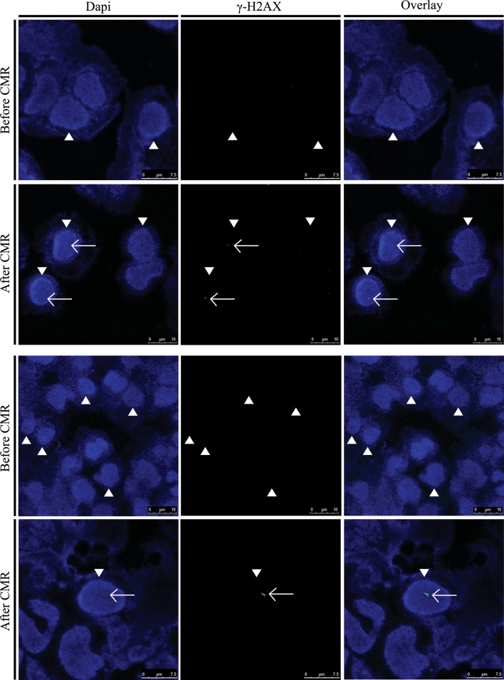
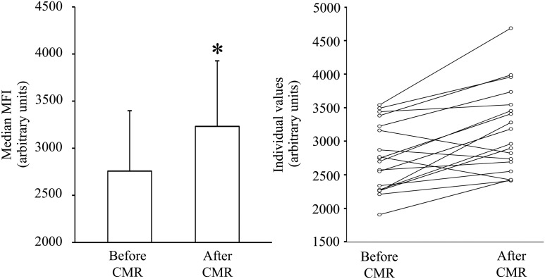
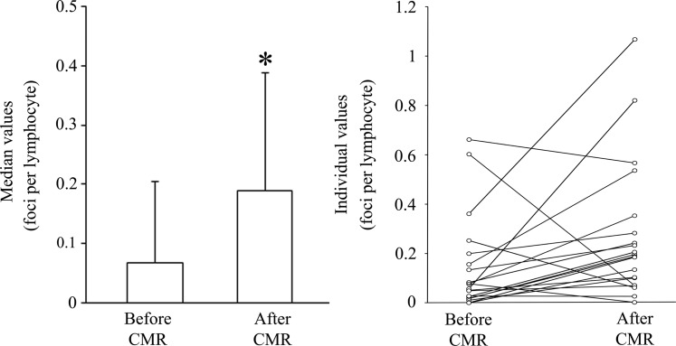
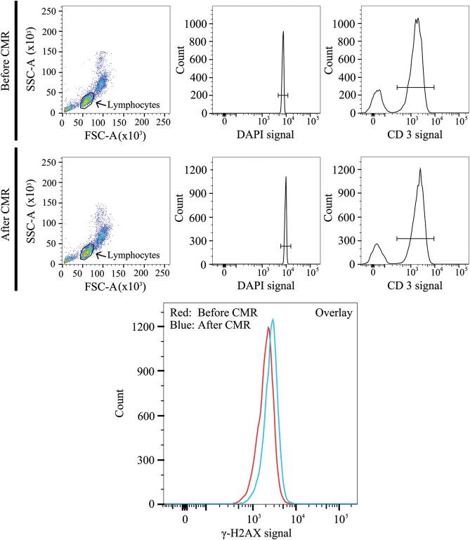
In 20 prospectively enrolled patients, peripheral venous blood was drawn before and after 1.5 T CMR scanning. After density gradient cell separation of blood samples, DNA DSBs in lymphocytes were quantified using immunofluorescence microscopy and flow cytometric analysis. Wilcoxon signed-rank testing was used for statistical analysis. Immunofluorescence microscopic and flow cytometric analysis revealed a significant increase in median numbers of DNA DSBs in lymphocytes induced by routine 1.5 T CMR examination.
The present findings indicate that CMR should be used with caution and that similar restrictions may apply as for X-ray-based and nuclear imaging techniques in order to avoid unnecessary damage of DNA integrity with potential carcinogenic effect.
磁共振(MR)成像在医学诊断成像中被广泛应用,因为它被认为是一种安全的替代电离辐射技术。最近有报道称,在心脏磁共振(CMR)中使用的强而快速切换的电磁场梯度可能具有潜在的遗传毒性效应,这引起了人们对安全性的关注。本研究旨在分析 CMR 检查前后人血淋巴细胞中的 DNA 双链断裂(DSB)。
在 20 例前瞻性入组的患者中,在 1.5 T CMR 扫描前后采集外周静脉血。在对血液样本进行密度梯度细胞分离后,使用免疫荧光显微镜和流式细胞术分析来定量淋巴细胞中的 DNA DSB。采用 Wilcoxon 符号秩检验进行统计学分析。免疫荧光显微镜和流式细胞术分析显示,常规 1.5 T CMR 检查可显著增加淋巴细胞中 DNA DSB 的中位数。
本研究结果表明,CMR 的使用应谨慎,并且可能需要与 X 射线和核成像技术类似的限制,以避免 DNA 完整性的不必要损伤,从而潜在地产生致癌作用。