Saraf Shyam K, Singh Ravindra P, Singh Vakil, Varma Ashish
Department of Orthopaedics, Institute of Medical Sciences, Banaras Hindu University, Varanasi, India.
Indian J Orthop. 2013 May;47(3):238-43. doi: 10.4103/0019-5413.111502.
The objective of this cadaveric study was to analyze the effects of iatrogenic pedicle perforations from screw misplacement on the mean pullout strength of lower thoracic and lumbar pedicle screws. We also investigated the effect of bone mineral density (BMD), diameter of pedicle screws, and the region of spine on the pullout strength of pedicle screws.
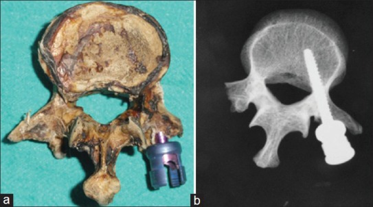
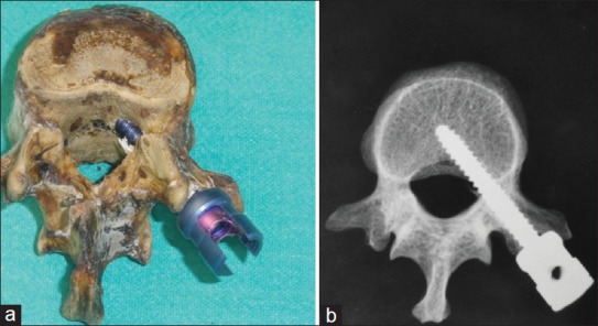
Sixty fresh human cadaveric vertebrae (D10-L2) were harvested. Dual-energy X-ray absorptiometry (DEXA) scan of vertebrae was done for BMD. Titanium pedicle screws of different diameters (5.2 and 6.2 mm) were inserted in the thoracic and lumbar segments after dividing the specimens into three groups: a) standard pedicle screw (no cortical perforation); b) screw with medial cortical perforation; and c) screw with lateral cortical perforation. Finally, pullout load of pedicle screws was recorded using INSTRON Universal Testing Machine.
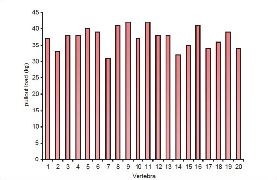
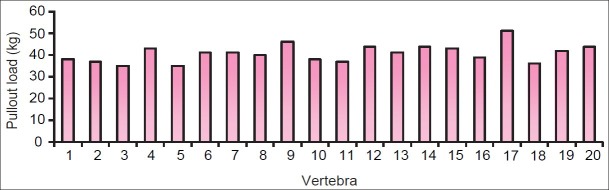
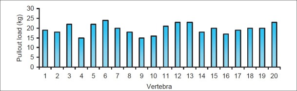
Compared with standard placement, medially misplaced screws had 9.4% greater mean pullout strength and laterally misplaced screws had 47.3% lesser mean pullout strength. The pullout strength of the 6.2 mm pedicle screws was 33% greater than that of the 5.2 mm pedicle screws. The pullout load of pedicle screws in lumbar vertebra was 13.9% greater than that in the thoracic vertebra (P = 0.105), but it was not statistically significant. There was no significant difference between pullout loads of vertebra with different BMD (P = 0.901).
The mean pullout strength was less with lateral misplaced pedicle screws while medial misplaced pedicle screw had more pullout strength. The pullout load of 6.2 mm screws was greater than that of 5.2 mm pedicle screws. No significant correlation was found between bone mineral densities and the pullout strength of vertebra. Similarly, the pullout load of screw placed in thoracic and lumbar vertebrae was not significantly different.
本尸体研究的目的是分析螺钉误置导致的医源性椎弓根穿孔对下胸椎和腰椎椎弓根螺钉平均拔出强度的影响。我们还研究了骨密度(BMD)、椎弓根螺钉直径和脊柱区域对椎弓根螺钉拔出强度的影响。
收集60个新鲜人尸体的椎骨(D10-L2)。对椎骨进行双能X线吸收法(DEXA)扫描以测定骨密度。将标本分为三组后,在胸段和腰段插入不同直径(5.2和6.2 mm)的钛制椎弓根螺钉:a)标准椎弓根螺钉(无皮质穿孔);b)内侧皮质穿孔的螺钉;c)外侧皮质穿孔的螺钉。最后,使用英斯特朗万能试验机记录椎弓根螺钉的拔出载荷。
与标准置入相比,内侧误置的螺钉平均拔出强度高9.4%,外侧误置的螺钉平均拔出强度低47.3%。6.2 mm椎弓根螺钉的拔出强度比5.2 mm椎弓根螺钉高33%。腰椎椎弓根螺钉的拔出载荷比胸椎高13.9%(P = 0.105),但无统计学意义。不同骨密度的椎骨拔出载荷之间无显著差异(P = 0.901)。
外侧误置的椎弓根螺钉平均拔出强度较低,而内侧误置的椎弓根螺钉拔出强度较高。6.2 mm螺钉的拔出载荷大于5.2 mm椎弓根螺钉。未发现骨密度与椎骨拔出强度之间存在显著相关性。同样,置于胸椎和腰椎的螺钉拔出载荷无显著差异。