Department of Virology II, National Institute of Infectious Diseases, Musashi-murayama, Tokyo, Japan.
PLoS One. 2013 Jun 14;8(6):e66534. doi: 10.1371/journal.pone.0066534. Print 2013.
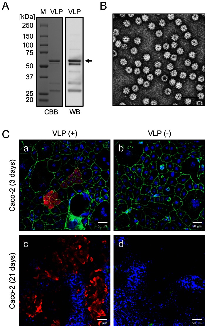
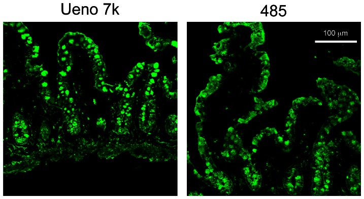
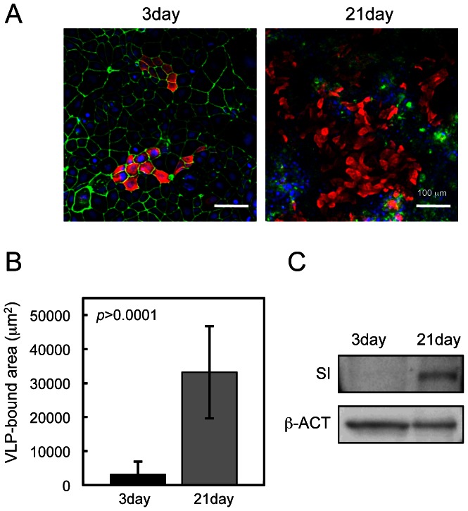
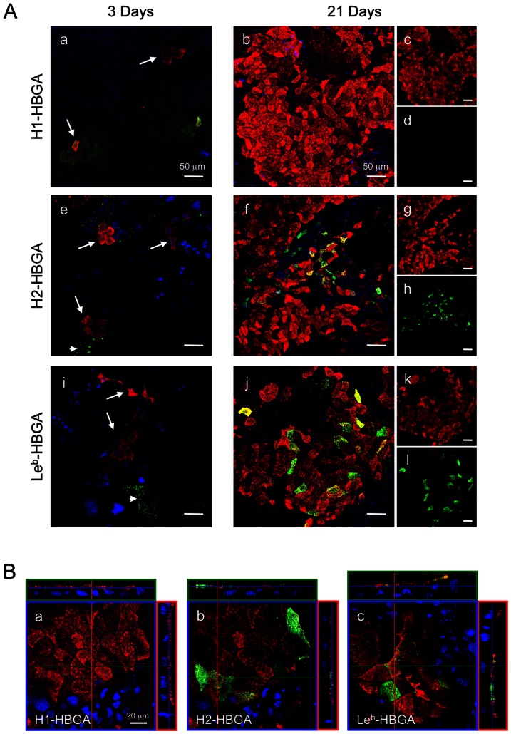
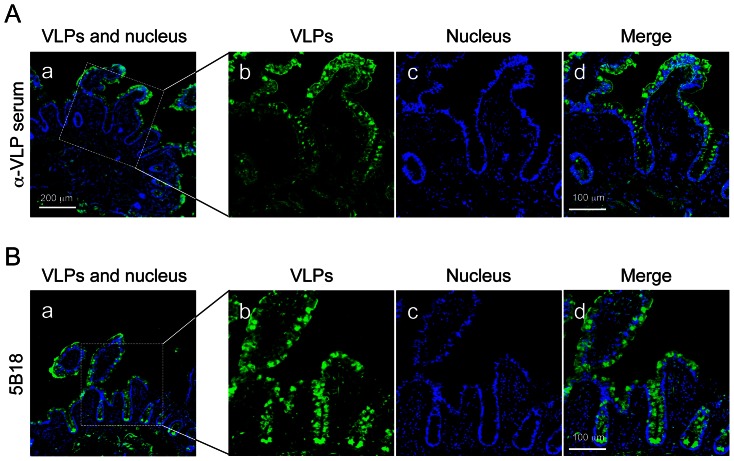
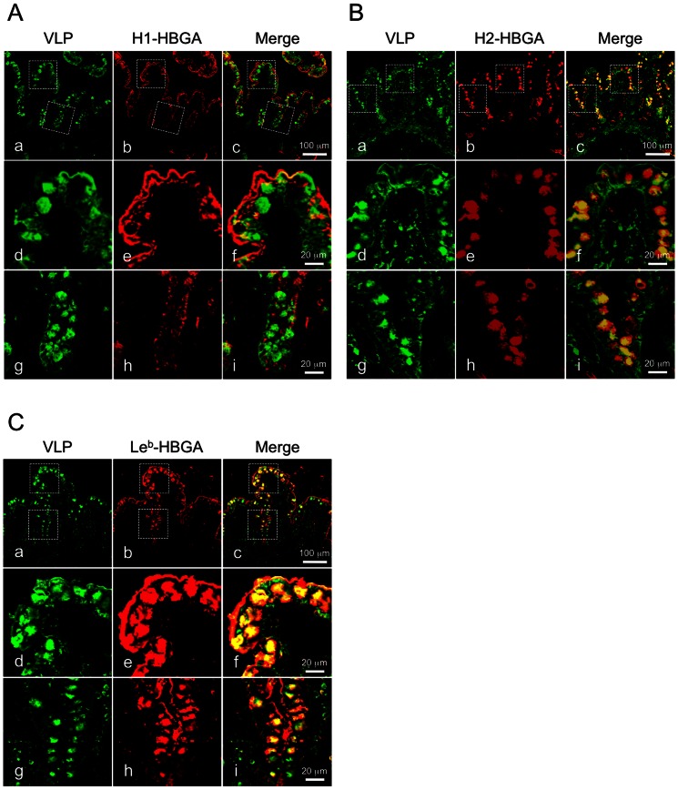
Human noroviruses (NoVs) are a major cause of non-bacterial gastroenteritis. Although histo-blood group antigens (HBGAs) have been implicated in the initial binding of NoV, the mechanism of that binding before internalization is not clear. To determine the involvement of NoVs and HBGAs in cell binding, we examined the localization of NoV virus-like particles (VLPs) and HBGAs in a human intestinal cell line and the human ileum biopsy specimens by immunofluorescence microscopy. The localizations of Ueno 7k VLPs (genogroup II.6) and each HBGA (type H1-, H2- and Le(b)-HBGAs) on the human intestinal cell line, Caco-2, were examined by confocal laser-scanning microscopy. To explore any interactions of NoVs and HBGAs in vivo, fresh biopsy specimens from human ileum were directly incubated with NoV VLPs and examined by immunofluorescence microscopy. We found that VLP binding depended on the state of cell differentiation, but not on the presence of HBGAs. In differentiated Caco-2 cells, we detected no type H1 HBGAs, but VLPs bound to the cells anyway. We incubated fresh biopsies of human ileum directly with VLPs, a model that better replicates the in vivo environment. VLPs mainly bound epithelial cells and goblet cells. Although the incubations were performed at 4°C to hinder internalization, VLPs were still detected inside cells. Our results suggest that VLPs utilize molecule(s) other than HBGAs during binding and internalization into cells.
人类诺如病毒(NoV)是导致非细菌性肠胃炎的主要原因。尽管组织血型抗原(HBGAs)已被认为与 NoV 的初始结合有关,但在内化之前,这种结合的机制尚不清楚。为了确定 NoV 和 HBGAs 在细胞结合中的作用,我们通过免疫荧光显微镜检查了 NoV 病毒样颗粒(VLPs)和 HBGAs 在人肠细胞系和人回肠活检标本中的定位。通过共聚焦激光扫描显微镜检查了 Ueno 7k VLPs(基因群 II.6)和每种 HBGA(H1-、H2-和 Le(b)-HBGAs)在人肠细胞系 Caco-2 上的定位。为了探索 NoV 和 HBGAs 在体内的任何相互作用,我们直接将新鲜的回肠活检标本与 NoV VLPs 孵育并用免疫荧光显微镜检查。我们发现 VLP 结合取决于细胞分化状态,但与 HBGAs 的存在无关。在分化的 Caco-2 细胞中,我们检测不到 H1 型 HBGAs,但 VLP 仍然与细胞结合。我们直接将人回肠的新鲜活检标本与 VLPs 孵育,这种模型更好地模拟了体内环境。VLPs 主要与上皮细胞和杯状细胞结合。尽管孵育在 4°C 下进行以阻止内化,但仍在细胞内检测到 VLPs。我们的结果表明,VLPs 在结合和内化到细胞中时利用了除 HBGAs 之外的分子。