Department of Pharmacology and Experimental Neuroscience, University of Nebraska Medical Center, Omaha, Nebraska, USA.
PLoS One. 2013 Jun 20;8(6):e66241. doi: 10.137/journal.pone.0066241. Print 2013.
Shortly after infection, HIV enters the brain and causes widespread inflammation and neuronal damage, which ultimately leads to neuropsychological impairments. Despite a large body of neuroscience and imaging studies, the pathophysiology of these HIV-associated neurocognitive disorders (HAND) remains unresolved. Previous neuroimaging studies have shown greater activation in HIV-infected patients during strenuous tasks in frontal and parietal cortices, and less activation in the primary sensory cortices during rest and sensory stimulation.
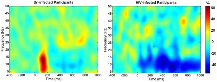
High-density magnetoencephalography (MEG) was utilized to evaluate the basic neurophysiology underlying attentive, visual processing in older HIV-infected adults and a matched non-infected control group. Unlike other neuroimaging methods, MEG is a direct measure of neural activity that is not tied to brain metabolism or hemodynamic responses. During MEG, participants fixated on a centrally-presented crosshair while intermittent visual stimulation appeared in their top-right visual-field quadrant. All MEG data was imaged in the time-frequency domain using beamforming.
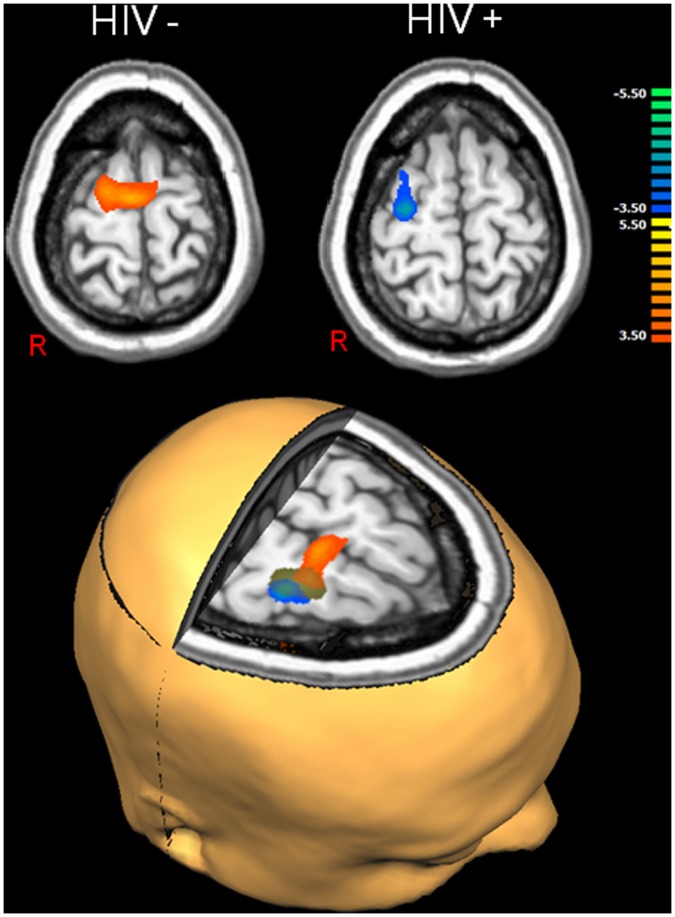
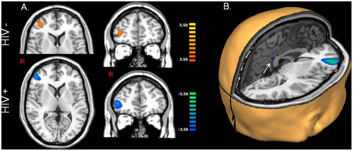
Uninfected controls had increased neuronal synchronization in the 6-12 Hz range within the right dorsolateral prefrontal cortex, right frontal eye-fields, and the posterior cingulate. Conversely, HIV-infected patients exhibited decreased synchrony in these same neural regions, and the magnitude of these decreases was correlated with neuropsychological performance in several cortical association regions.
MEG-based imaging holds potential as a noninvasive biomarker for HIV-related neuronal dysfunction, and may help identify patients who have or may develop HAND. Reduced synchronization of neural populations in the association cortices was strongly linked to cognitive dysfunction, and likely reflects the impact of HIV on neuronal and neuropsychological health.
HIV 感染后不久,便会进入大脑,引发广泛的炎症和神经元损伤,最终导致神经心理障碍。尽管有大量神经科学和影像学研究,但这些与 HIV 相关的神经认知障碍(HAND)的病理生理学仍未得到解决。先前的神经影像学研究表明,HIV 感染患者在进行剧烈任务时,前额叶和顶叶皮质的活跃度更高,而在休息和感觉刺激时,初级感觉皮质的活跃度更低。
利用高密度脑磁图(MEG)评估老年 HIV 感染患者和匹配的未感染对照组在注意力、视觉处理方面的基本神经生理学。与其他神经影像学方法不同,MEG 是一种直接测量神经活动的方法,不受大脑代谢或血液动力学反应的影响。在 MEG 期间,参与者注视中央呈现的十字准线,同时间歇性地在右上视野象限呈现视觉刺激。所有 MEG 数据都使用波束形成在时频域中进行成像。
未感染的对照组在右侧背外侧前额叶皮质、右侧额眼区和后扣带回的 6-12 Hz 范围内显示出神经元同步性增加。相反,HIV 感染患者在这些相同的神经区域显示出同步性降低,并且这些降低的幅度与几个皮质联合区域的神经心理学表现相关。
基于 MEG 的成像具有作为 HIV 相关神经元功能障碍的非侵入性生物标志物的潜力,并且可能有助于识别患有或可能患有 HAND 的患者。联合皮质中神经元群体同步性的降低与认知功能障碍密切相关,可能反映了 HIV 对神经元和神经心理健康的影响。