Department of Radiology, Shanghai Renji Hospital, Shanghai Jiao Tong University School of Medicine, Shanghai, China.
PLoS One. 2013 Jul 5;8(7):e69701. doi: 10.1371/journal.pone.0069701. Print 2013.
To investigate the feasibility of gadolinium (Gd) contrast-enhanced magnetic resonance lymphangiography (MRL) in breast cancer patients within a typical clinical setting, and to establish a Gd-MRL protocol and identify potential MRL biomarkers for differentiating metastatic from non-metastatic lymph nodes.
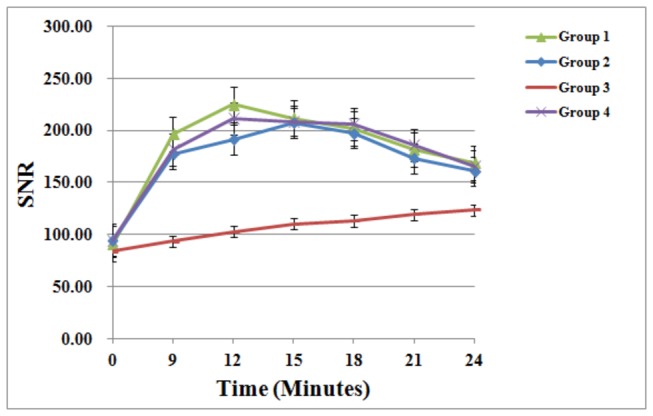
32 patients with unilateral breast cancer were enrolled and divided into 4 groups of 8 patients. Groups I, II, and III received 1.0, 0.5, and 0.3 ml of intradermal contrast; group IV received two 0.5 ml doses of intradermal contrast. MRL images were acquired on a 3.0 T system and evaluated independently by two radiologists for the number and size of enhancing lymph nodes, lymph node contrast uptake kinetics, lymph vessel size, and contrast enhancement patterns within lymph nodes.
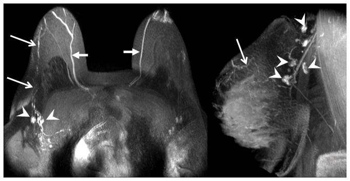
Group III patients had a statistically significant decrease in the total number of enhancing axillary lymph nodes and lymphatic vessels compared to all other groups. While group IV patients had a statistically significant faster time to reach the maximum peak enhancement over group I and II (by 3 minutes), there was no other statistically significant difference between imaging results between groups I, II, and IV. 27 out of 128 lymphatic vessels (21%) showed dilatation, and all patients with dilated lymphatic vessels were pathologically proven to have metastases. Using the pattern of enhancement defects as the sole criterion for identifying metastatic lymph nodes during Gd-MRL interpretation, and using histopathology as the gold standard, the sensitivity and specificity were estimated to be 86% and 95%, respectively.
Gd-MRL can adequately depict the lymphatic system, can define sentinel lymph nodes, and has the potential to differentiate between metastatic and non-metastatic lymph nodes in breast cancer patients.
在典型的临床环境下,研究钆(Gd)增强磁共振淋巴造影(MRL)在乳腺癌患者中的可行性,并建立 Gd-MRL 方案,确定潜在的 MRL 生物标志物,以区分转移性和非转移性淋巴结。
共纳入 32 例单侧乳腺癌患者,分为 4 组,每组 8 例。I、II、III 组分别接受 1.0、0.5 和 0.3 ml 皮内对比剂;IV 组接受两次 0.5 ml 皮内对比剂。使用 3.0 T 系统采集 MRL 图像,由 2 名放射科医生独立评估增强淋巴结的数量和大小、淋巴结对比摄取动力学、淋巴管大小以及淋巴结内的对比增强模式。
与其他组相比,III 组患者腋窝增强淋巴结和淋巴管总数明显减少。虽然 IV 组患者达到最大峰值增强的时间比 I 组和 II 组分别提前 3 分钟(具有统计学意义),但各组间的影像学结果无其他统计学差异。128 条淋巴管中的 27 条(21%)出现扩张,所有扩张淋巴管患者均经病理证实存在转移。在 Gd-MRL 解读中,仅将增强缺陷模式作为识别转移性淋巴结的唯一标准,以组织病理学为金标准,其敏感性和特异性估计分别为 86%和 95%。
Gd-MRL 可充分显示淋巴系统,能够定位前哨淋巴结,并有可能区分乳腺癌患者的转移性和非转移性淋巴结。