Dissaranan Charuspong, Cruz Michelle A, Kiedrowski Matthew J, Balog Brian M, Gill Bradley C, Penn Marc S, Goldman Howard B, Damaser Margot S
Glickman Urologic and Kidney Institute, Cleveland Clinic, Cleveland, OH, USA.
Cell Transplant. 2014;23(11):1395-406. doi: 10.3727/096368913X670921. Epub 2013 Jul 17.
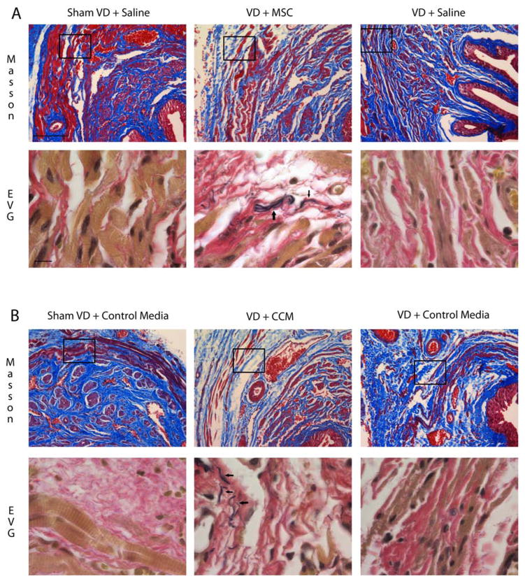
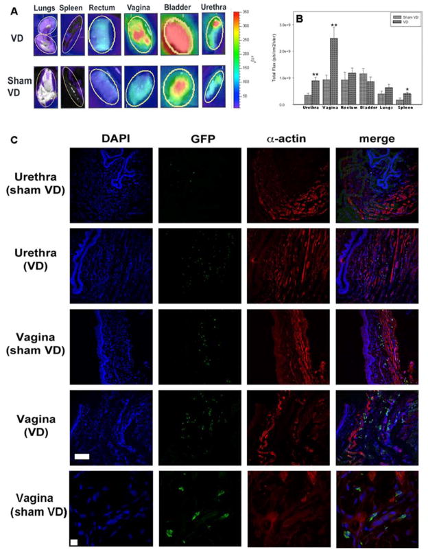
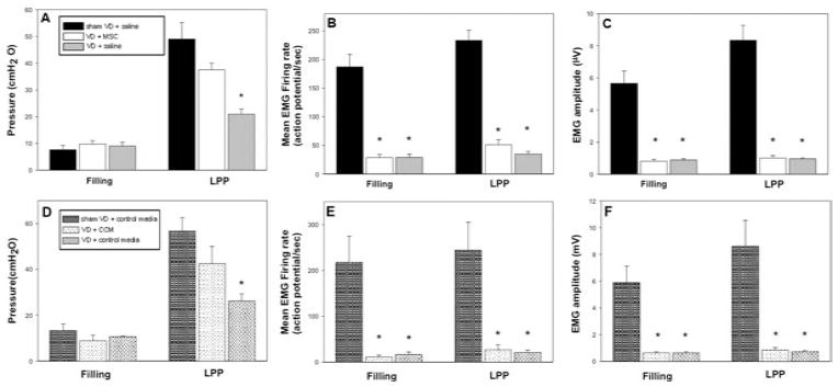
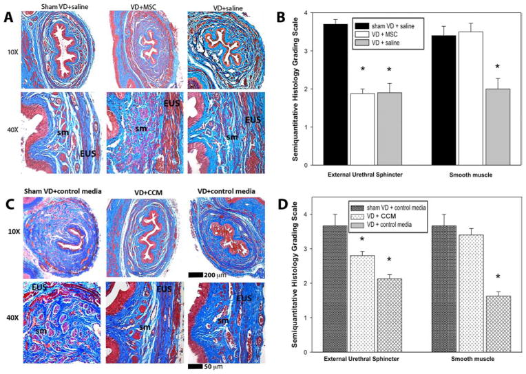
Vaginal delivery is a risk factor for stress urinary incontinence (SUI). Mesenchymal stem cells (MSCs) home to injured organs and can facilitate repair. The goal of this study was to determine if MSCs home to pelvic organs after simulated childbirth injury and facilitate recovery from SUI via paracrine factors. Three experiments were performed. Eighteen female rats received vaginal distension (VD) or sham VD and labeled intravenous (IV) MSCs to investigate if MSCs home to the pelvic organs. Whole-organ imaging and immunofluorescence were performed 1 week later. Thirty-four female rats received VD and IV MSCs, VD and IV saline, or sham VD and IV saline to investigate if MSCs accelerate recovery of continence. Twenty-nine female rats received VD and periurethral concentrated conditioned media (CCM), VD and periurethral control media, or sham VD and periurethral control media to investigate if factors secreted by MSCs accelerate recovery from VD. Urethral histology and function were assessed 1 week later. Significantly more MSCs were observed in the urethra, vagina, and spleen after VD compared to sham VD. Continence as measured by leak point pressure (LPP) was significantly reduced after VD in rats treated with saline or control media compared to sham VD but not in those given MSCs or CCM. External urethral sphincter (EUS) function as measured by electromyography (EMG) was not improved with MSCs or CCM. Rats treated with MSCs or CCM demonstrated an increase in elastin fibers near the EUS and urethral smooth muscle more similar to that of sham-injured animals than rats treated with saline or control media. MSCs homed to the urethra and vagina and facilitated recovery of continence most likely via secretion of paracrine factors. Both MSCs and CCM have promise as novel noninvasive therapies for SUI.
阴道分娩是压力性尿失禁(SUI)的一个风险因素。间充质干细胞(MSCs)会归巢至受损器官并能促进修复。本研究的目的是确定在模拟分娩损伤后MSCs是否会归巢至盆腔器官,并通过旁分泌因子促进SUI的恢复。进行了三项实验。18只雌性大鼠接受阴道扩张(VD)或假阴道扩张,并标记静脉注射(IV)的MSCs,以研究MSCs是否归巢至盆腔器官。1周后进行全器官成像和免疫荧光检测。34只雌性大鼠接受VD和IV MSCs、VD和IV生理盐水或假阴道扩张和IV生理盐水,以研究MSCs是否能加速控尿功能的恢复。29只雌性大鼠接受VD和尿道周围浓缩条件培养基(CCM)、VD和尿道周围对照培养基或假阴道扩张和尿道周围对照培养基,以研究MSCs分泌的因子是否能加速VD后的恢复。1周后评估尿道组织学和功能。与假阴道扩张相比,VD后在尿道、阴道和脾脏中观察到的MSCs明显更多。与假阴道扩张相比,用生理盐水或对照培养基处理的大鼠在VD后通过漏点压力(LPP)测量的控尿功能明显降低,但给予MSCs或CCM的大鼠则没有。通过肌电图(EMG)测量的尿道外括约肌(EUS)功能在给予MSCs或CCM后没有改善。与用生理盐水或对照培养基处理的大鼠相比,用MSCs或CCM处理的大鼠在EUS和尿道平滑肌附近的弹性纤维增加,更类似于假损伤动物。MSCs归巢至尿道和阴道,并最有可能通过旁分泌因子的分泌促进控尿功能的恢复。MSCs和CCM都有望成为治疗SUI的新型非侵入性疗法。