Support Center for Advanced Neuroimaging, University Institute of Diagnostic and Interventional Neuroradiology, Inselspital, University of Berne, Bern, Switzerland.
PLoS One. 2013 Jul 16;8(7):e67610. doi: 10.1371/journal.pone.0067610. Print 2013.
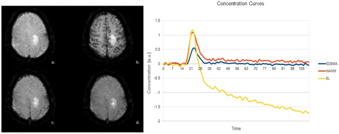
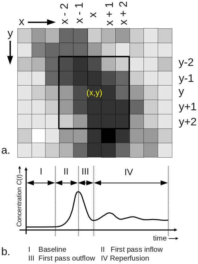
Texture analysis is an alternative method to quantitatively assess MR-images. In this study, we introduce dynamic texture parameter analysis (DTPA), a novel technique to investigate the temporal evolution of texture parameters using dynamic susceptibility contrast enhanced (DSCE) imaging. Here, we aim to introduce the method and its application on enhancing lesions (EL), non-enhancing lesions (NEL) and normal appearing white matter (NAWM) in multiple sclerosis (MS).
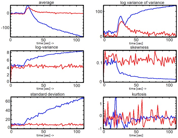
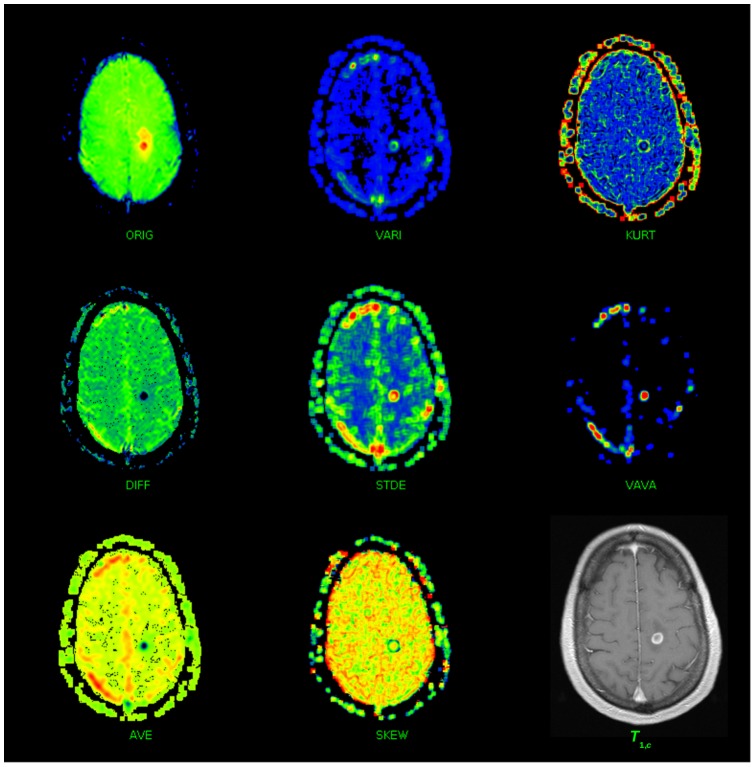
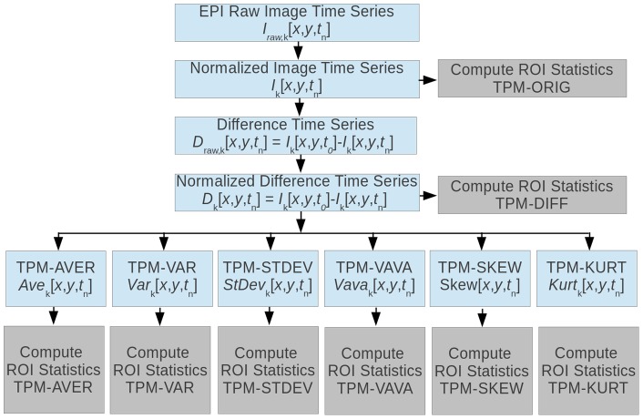
We investigated 18 patients with MS and clinical isolated syndrome (CIS), according to the 2010 McDonald's criteria using DSCE imaging at different field strengths (1.5 and 3 Tesla). Tissues of interest (TOIs) were defined within 27 EL, 29 NEL and 37 NAWM areas after normalization and eight histogram-based texture parameter maps (TPMs) were computed. TPMs quantify the heterogeneity of the TOI. For every TOI, the average, variance, skewness, kurtosis and variance-of-the-variance statistical parameters were calculated. These TOI parameters were further analyzed using one-way ANOVA followed by multiple Wilcoxon sum rank testing corrected for multiple comparisons.
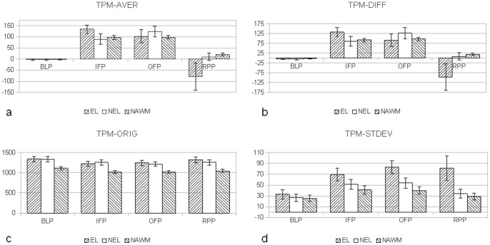
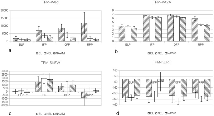
Tissue- and time-dependent differences were observed in the dynamics of computed texture parameters. Sixteen parameters discriminated between EL, NEL and NAWM (pAVG = 0.0005). Significant differences in the DTPA texture maps were found during inflow (52 parameters), outflow (40 parameters) and reperfusion (62 parameters). The strongest discriminators among the TPMs were observed in the variance-related parameters, while skewness and kurtosis TPMs were in general less sensitive to detect differences between the tissues.
DTPA of DSCE image time series revealed characteristic time responses for ELs, NELs and NAWM. This may be further used for a refined quantitative grading of MS lesions during their evolution from acute to chronic state. DTPA discriminates lesions beyond features of enhancement or T2-hypersignal, on a numeric scale allowing for a more subtle grading of MS-lesions.
纹理分析是一种定量评估磁共振图像的替代方法。本研究引入动态纹理参数分析(DTPA),这是一种使用动态对比增强磁共振成像(DSCE)研究纹理参数时间演变的新技术。在这里,我们旨在介绍该方法及其在多发性硬化症(MS)中增强病变(EL)、非增强病变(NEL)和正常表现的白质(NAWM)中的应用。
根据 2010 年麦克唐纳标准,我们对 18 名 MS 患者和临床孤立综合征(CIS)患者进行了研究,在不同场强(1.5 和 3 Tesla)下进行了 DSCE 成像。在归一化后,在 27 个 EL、29 个 NEL 和 37 个 NAWM 区域内定义感兴趣的组织(TOI),并计算了 8 个基于直方图的纹理参数图(TPM)。TPM 量化了 TOI 的异质性。对于每个 TOI,计算了平均值、方差、偏度、峰度和方差的方差统计参数。对这些 TOI 参数进行了单向方差分析,然后进行了多个威尔科克森符号秩检验,并用多重比较校正。
观察到计算的纹理参数在组织和时间上存在依赖性差异。16 个参数可区分 EL、NEL 和 NAWM(pAVG=0.0005)。在流入(52 个参数)、流出(40 个参数)和再灌注(62 个参数)期间,在 DTPA 纹理图中发现了显著差异。在 TPM 中,方差相关参数是最强的鉴别器,而偏度和峰度 TPM 通常对检测组织之间的差异不太敏感。
DSCE 图像时间序列的 DTPA 显示出 EL、NEL 和 NAWM 的特征时间反应。这可能进一步用于在 MS 病变从急性到慢性状态的演变过程中对其进行更精细的定量分级。DTPA 可在数字尺度上区分病变,超越增强或 T2 高信号的特征,从而对 MS 病变进行更细微的分级。