The Department of Plastic Surgery, The University of Texas MD Anderson Cancer Center, Houston, Texas, United States of America.
PLoS One. 2013 Jul 24;8(7):e69222. doi: 10.1371/journal.pone.0069222. Print 2013.
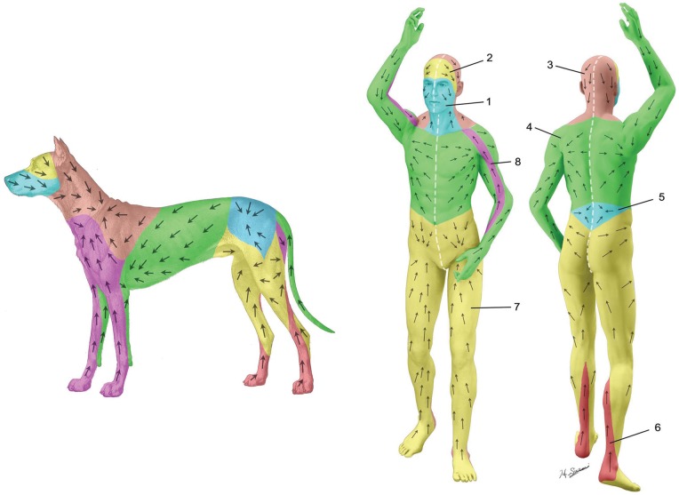
Lymph node dissection is often performed as a part of surgical treatment for breast cancer and malignant melanoma to prevent malignant cells from traveling via the lymphatic system. Currently little is known about postoperative lymphatic drainage pattern alterations. This knowledge may be useful for management of recurrent cancer and prevention of breast cancer related lymphedema. We mapped the complete superficial lymphatic system of a dog and used this canine model to perform preliminary studies of lymphatic architectural changes in postoperative condition.
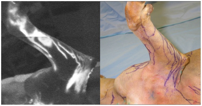
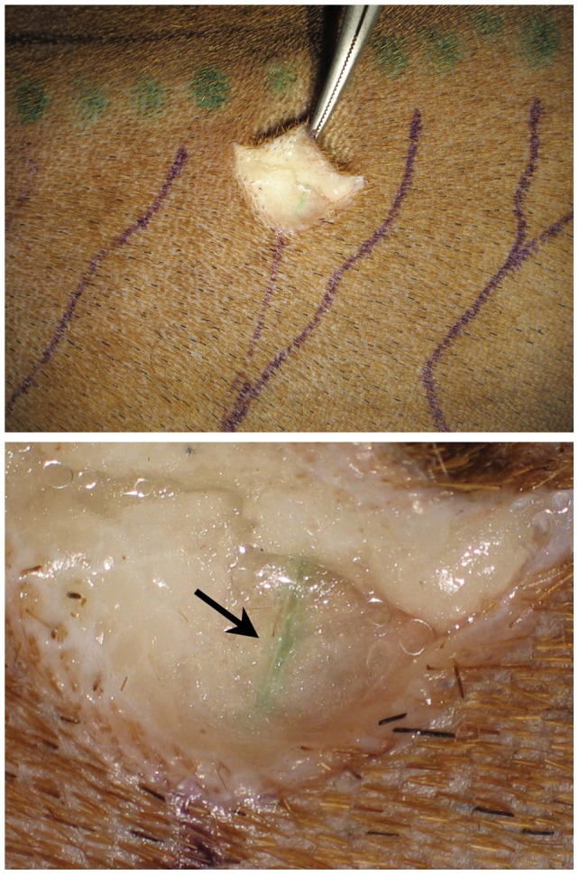
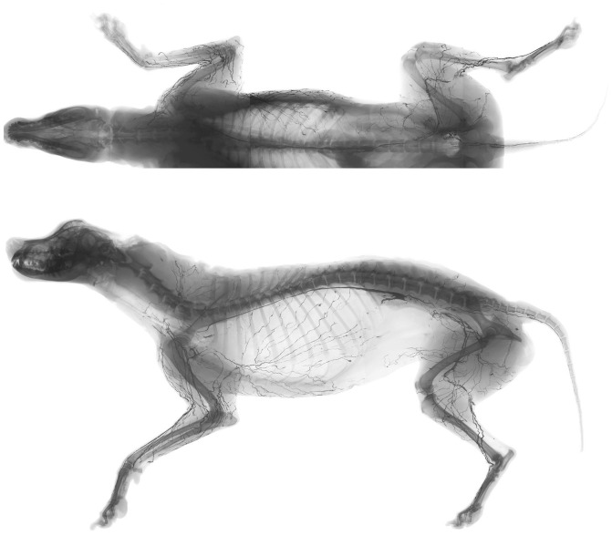
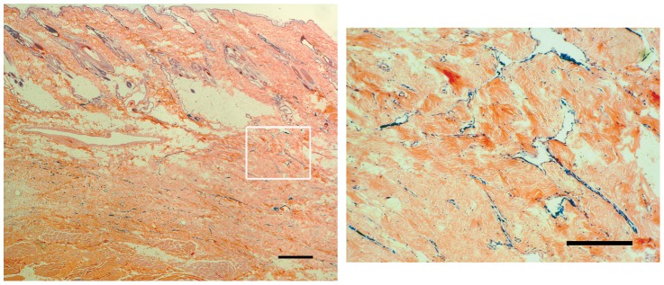
Lymphatic territories (lymphosomes) were mapped with 4 female mongrel carcasses using an indocyanine green (ICG) fluorescent lymphography and a radiographic microinjection technique. Two live dogs were then subjected to unilateral lymph node dissection of lymph basins of the forelimb, and ICG lymphography and lymphangiogram were performed 6 months after the surgery to investigate lymphatic changes. Lymphatic patterns in the carcass were then compared with postoperative lymphatic patterns in the live dogs.
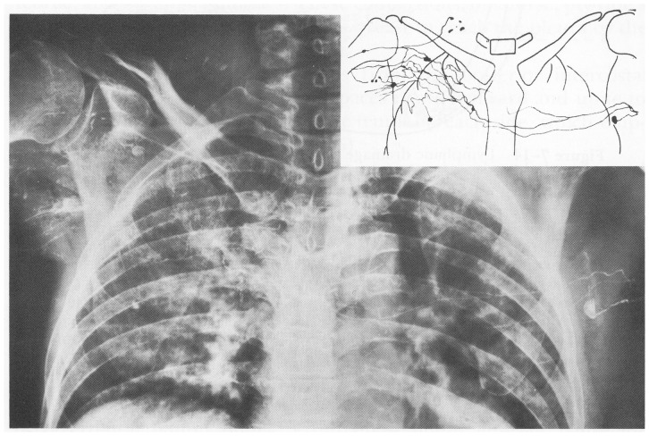
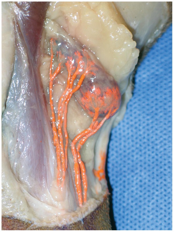
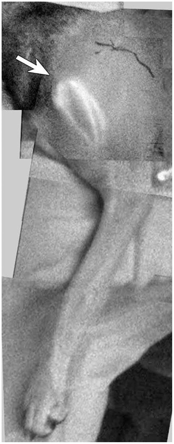
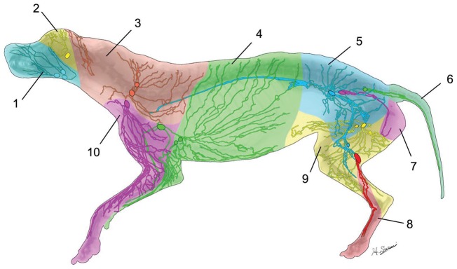
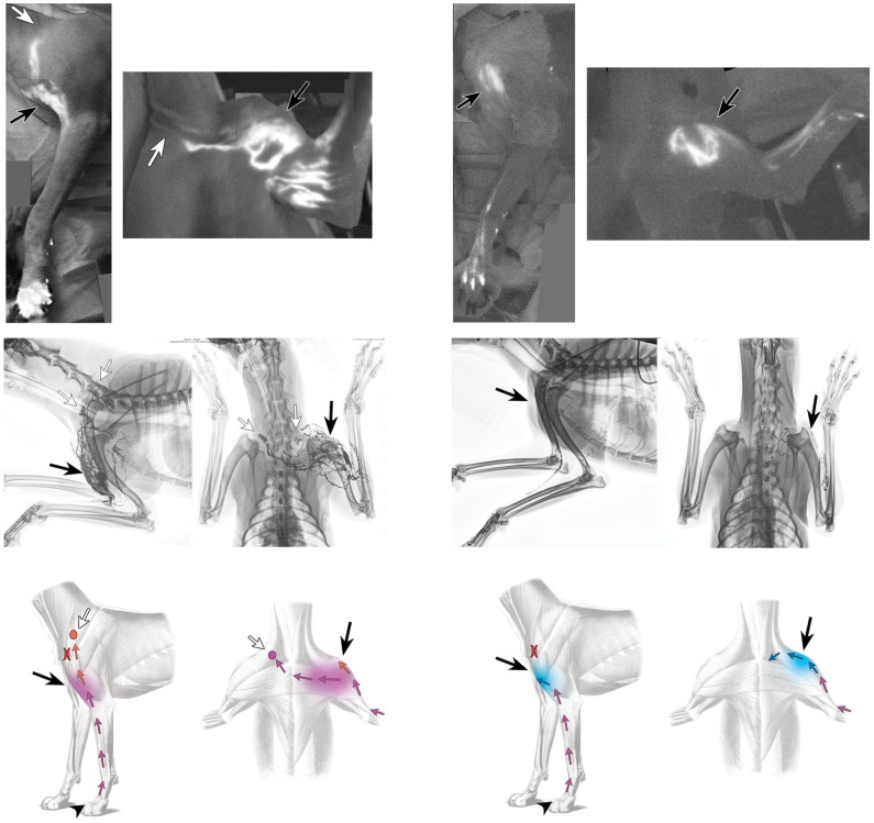
Ten lymphosomes were identified, corresponding with ten lymphatic basins. Postoperative fluorescent lymphographic images and lymphangiograms in the live dogs revealed small caliber lymphatic network fulfilling gaps in the surgical area and collateral lymphatic vessels arising from the network connecting to lymph nodes in the contralateral and ipsilateral neck in one dog and the ipsilateral subclavicular vein in another dog.
Our canine lymphosome map allowed us to observe lymphatic collateral formations after lymph node dissection in live dogs. This canine model may help clarify our understanding of postoperative lymphatic changes in humans in future studies.
淋巴结清扫术常作为乳腺癌和恶性黑色素瘤手术治疗的一部分,以防止恶性细胞通过淋巴系统转移。目前,人们对术后淋巴引流模式的改变知之甚少。这些知识对于管理复发性癌症和预防乳腺癌相关的淋巴水肿可能是有用的。我们描绘了一只狗的完整浅层淋巴系统,并使用这种犬模型对术后淋巴结构变化进行了初步研究。
使用吲哚菁绿(ICG)荧光淋巴造影和放射性微注射技术,对 4 只雌性杂种犬尸体的淋巴区(淋巴管)进行了定位。然后对 2 只活狗进行了单侧前肢淋巴结清扫,并在手术后 6 个月进行了 ICG 淋巴造影和淋巴管造影,以研究淋巴变化。然后将尸体的淋巴模式与活狗的术后淋巴模式进行比较。
确定了 10 个淋巴管,对应 10 个淋巴区。在活狗中,术后荧光淋巴造影图像和淋巴管造影显示,小口径淋巴管网络填补了手术区域的空白,并且从网络中出现的侧支淋巴管连接到一只狗的对侧和同侧颈部淋巴结,另一只狗的同侧锁骨下静脉的淋巴结。
我们的犬淋巴管图谱使我们能够在活狗中观察到淋巴结清扫术后的侧支淋巴管形成。这种犬模型可能有助于在未来的研究中阐明我们对人类术后淋巴变化的理解。