Suami Hiroo, Scaglioni Mario F
Sydney, New South Wales, Australia; Houston, Texas; and Zurich, Switzerland.
From Australian Lymphoedema Education, Research and Treatment, Faculty of Medicine and Health Sciences, Macquarie University; the Department of Plastic Surgery, University of Texas M. D. Anderson Cancer Center; and the Department of Plastic Surgery and Hand Surgery, University Hospital Zurich.
Plast Reconstr Surg. 2017 Nov;140(5):945-951. doi: 10.1097/PRS.0000000000003776.
Understanding the precise anatomy in experimental animals is crucial for correct design of research projects. Rats are commonly used for scientific research in plastic surgery because of their availability in academic institutions, moderate cost, and sizable vessels for microsurgical procedures. In past publications about rat anatomy, lymphatic mapping has been limited and incomplete. The aim of this study was to comprehensively map the superficial lymphatic system in the rat.
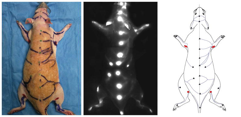
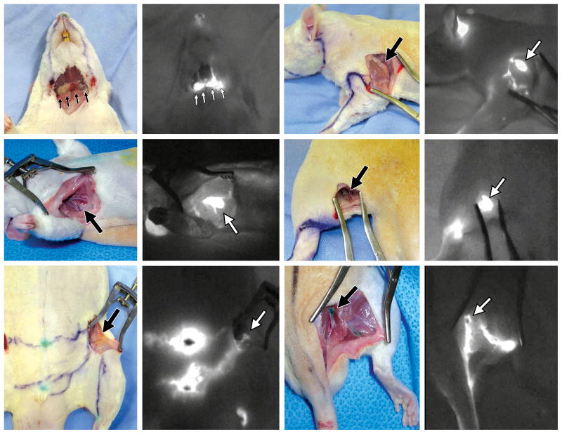
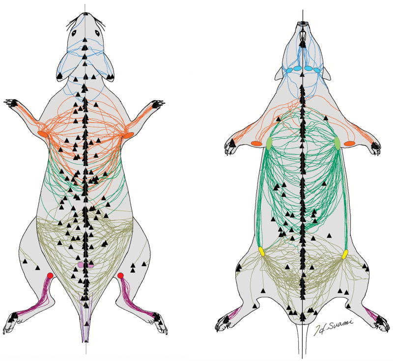
Twenty-seven Sprague-Dawley rats were used for this study. Indocyanine green fluorescence lymphography was used to identify the lymphatic vessels and lymph nodes. Under general anaesthesia, indocyanine green was injected intradermally at multiple spots along the dorsal and medial midlines, front and hind paws, ears, and tail. The course of the lymphatic vessels was traced on the skin with a marker pen and photographed. The superficial lymphatic vessels in each rat were sketched on a graphic template and all of the templates were superimposed using graphics software to define the relationship between the lymphatic vessel and sentinel node.
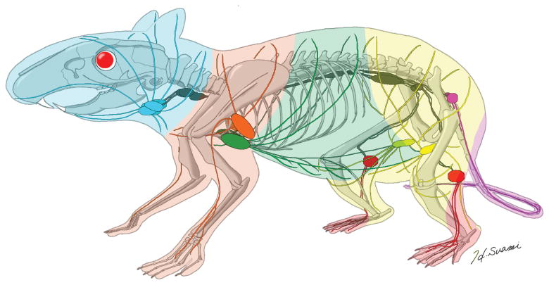
Indocyanine green fluorescence lymphography was able to demonstrate the superficial lymphatic vessels in the rat. Six groups of regional lymph node/s were identified and lymphatic pathways to those nodes delineated. The authors' lymphosome concept was successfully applied to the rat, with six lymphosomes identified.
The authors succeeded in performing superficial lymphatic mapping in the rat. The authors' anatomical findings can provide further information about the lymphatic system in the normal state and promote understanding of pathologic changes generated by surgical manipulation for future studies.
了解实验动物的确切解剖结构对于正确设计研究项目至关重要。大鼠因其在学术机构中易于获取、成本适中且有适合显微外科手术的较大血管,常用于整形外科的科学研究。在过去关于大鼠解剖学的出版物中,淋巴绘图有限且不完整。本研究的目的是全面绘制大鼠的浅表淋巴系统。
本研究使用了27只Sprague-Dawley大鼠。采用吲哚菁绿荧光淋巴造影术来识别淋巴管和淋巴结。在全身麻醉下,将吲哚菁绿多点皮内注射到沿背侧和内侧中线、前爪和后爪、耳朵及尾巴。用记号笔在皮肤上追踪淋巴管的走行并拍照。将每只大鼠的浅表淋巴管绘制在图形模板上,并使用图形软件将所有模板叠加,以确定淋巴管与前哨淋巴结之间的关系。
吲哚菁绿荧光淋巴造影术能够显示大鼠的浅表淋巴管。识别出六组区域淋巴结,并描绘了通向这些淋巴结的淋巴途径。作者的淋巴体概念成功应用于大鼠,识别出六个淋巴体。
作者成功地对大鼠进行了浅表淋巴绘图。作者的解剖学发现可为正常状态下的淋巴系统提供更多信息,并促进对未来研究中手术操作所产生病理变化的理解。