• 文献检索
  • 文档翻译
  • 深度研究
  • 学术资讯
  • Suppr Zotero 插件Zotero 插件
  • 邀请有礼
  • 套餐&价格
  • 历史记录
应用&插件
Suppr Zotero 插件Zotero 插件浏览器插件Mac 客户端Windows 客户端微信小程序
定价
高级版会员购买积分包购买API积分包
服务
文献检索文档翻译深度研究API 文档MCP 服务
关于我们
关于 Suppr公司介绍联系我们用户协议隐私条款
关注我们

Suppr 超能文献

核心技术专利:CN118964589B侵权必究
粤ICP备2023148730 号-1Suppr @ 2026

文献检索

告别复杂PubMed语法,用中文像聊天一样搜索,搜遍4000万医学文献。AI智能推荐,让科研检索更轻松。

立即免费搜索

文件翻译

保留排版,准确专业,支持PDF/Word/PPT等文件格式,支持 12+语言互译。

免费翻译文档

深度研究

AI帮你快速写综述,25分钟生成高质量综述,智能提取关键信息,辅助科研写作。

立即免费体验

窥视人类肾草酸钙结石基质:参与尿石症复杂机制的新型蛋白质的特征。

Peeping into human renal calcium oxalate stone matrix: characterization of novel proteins involved in the intricate mechanism of urolithiasis.

机构信息

Department of Biotechnology and Bioinformatics, Jaypee University of Information Technology, Waknaghat, Solan, Himachal Pradesh, India.

出版信息

PLoS One. 2013 Jul 24;8(7):e69916. doi: 10.1371/journal.pone.0069916. Print 2013.

DOI:10.1371/journal.pone.0069916
PMID:23894559
原文链接:https://pmc.ncbi.nlm.nih.gov/articles/PMC3722206/
Abstract

BACKGROUND

The increasing number of patients suffering from urolithiasis represents one of the major challenges which nephrologists face worldwide today. For enhancing therapeutic outcomes of this disease, the pathogenic basis for the formation of renal stones is the need of hour. Proteins are found as major component in human renal stone matrix and are considered to have a potential role in crystal-membrane interaction, crystal growth and stone formation but their role in urolithiasis still remains obscure.

METHODS

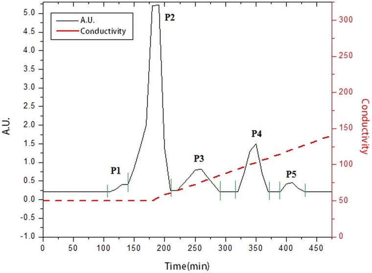
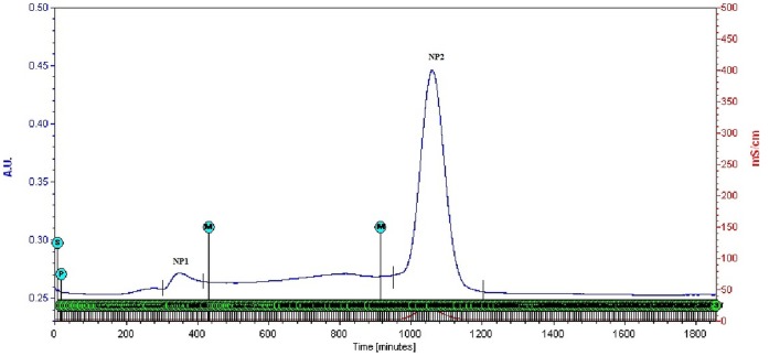
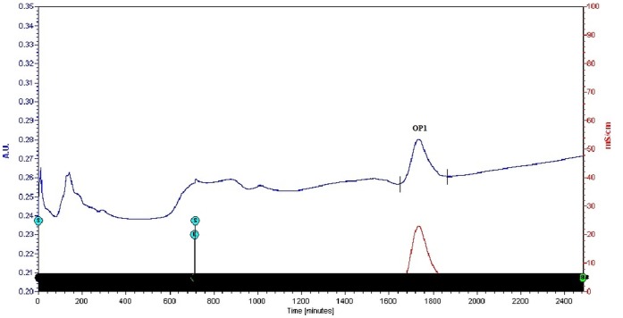
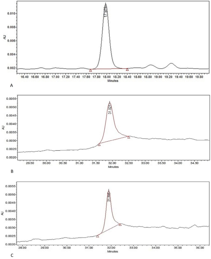
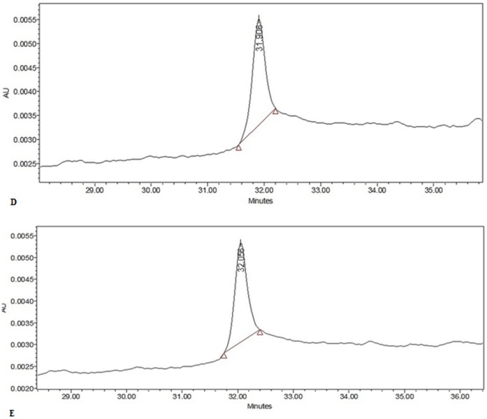
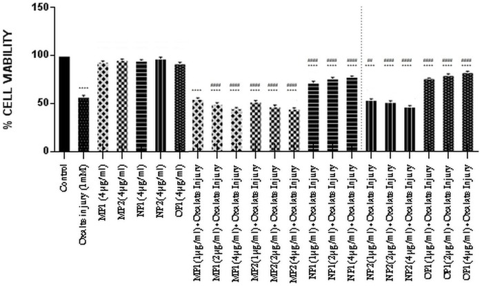
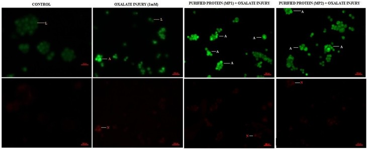
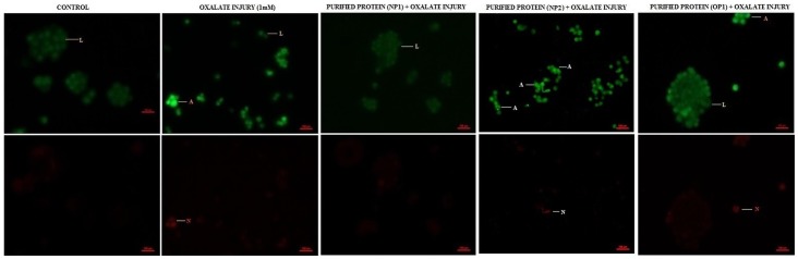
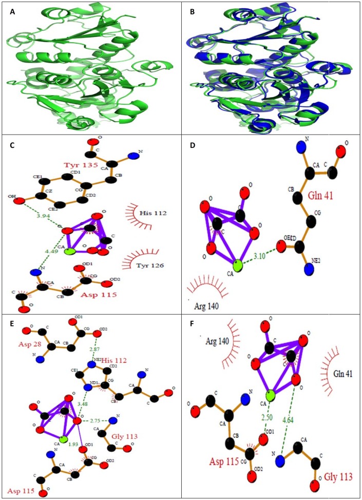
Proteins were isolated from the matrix of human CaOx containing kidney stones. Proteins having MW>3 kDa were subjected to anion exchange chromatography followed by molecular-sieve chromatography. The effect of these purified proteins was tested against CaOx nucleation and growth and on oxalate injured Madin-Darby Canine Kidney (MDCK) renal epithelial cells for their activity. Proteins were identified by Matrix-assisted laser desorption/ionization-time of flight (MALDI-TOF MS) followed by database search with MASCOT server. In silico molecular interaction studies with CaOx crystals were also investigated.

RESULTS

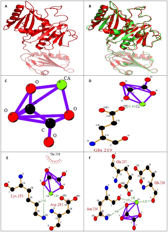
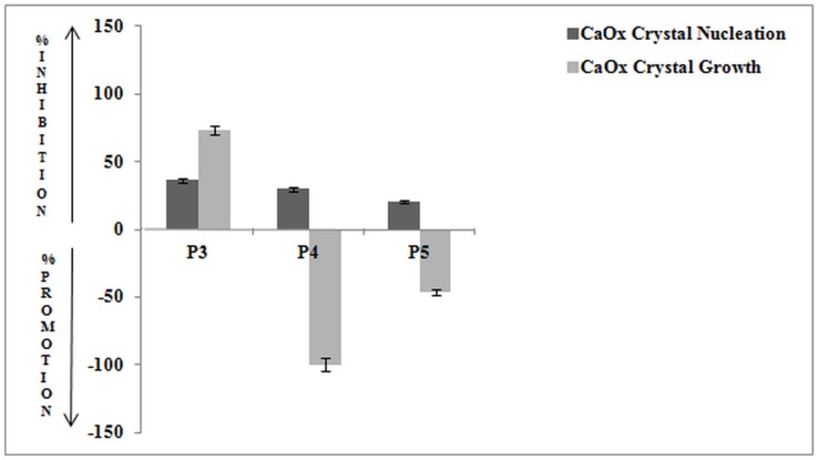
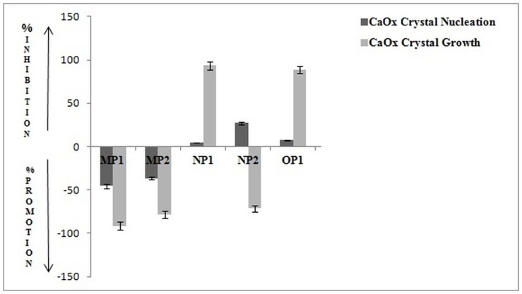
Five proteins were identified from the matrix of calcium oxalate kidney stones by MALDI-TOF MS followed by database search with MASCOT server with the competence to control the stone formation process. Out of which two proteins were promoters, two were inhibitors and one protein had a dual activity of both inhibition and promotion towards CaOx nucleation and growth. Further molecular modelling calculations revealed the mode of interaction of these proteins with CaOx at the molecular level.

CONCLUSIONS

We identified and characterized Ethanolamine-phosphate cytidylyltransferase, Ras GTPase-activating-like protein, UDP-glucose:glycoprotein glucosyltransferase 2, RIMS-binding protein 3A, Macrophage-capping protein as novel proteins from the matrix of human calcium oxalate stone which play a critical role in kidney stone formation. Thus, these proteins having potential to modulate calcium oxalate crystallization will throw light on understanding and controlling urolithiasis in humans.

摘要

背景

越来越多的尿路结石患者是当今全球肾病学家面临的主要挑战之一。为了提高这种疾病的治疗效果,肾结石形成的病理基础是当务之急。蛋白质是人类肾石基质的主要成分,被认为在晶体-膜相互作用、晶体生长和结石形成中具有潜在作用,但它们在尿石症中的作用仍不清楚。

方法

从人草酸钙结石基质中分离出蛋白质。MW>3 kDa 的蛋白质进行阴离子交换层析,然后进行分子筛层析。测试这些纯化蛋白对草酸钙成核和生长的影响,以及对草酸损伤的 Madin-Darby Canine Kidney (MDCK) 肾上皮细胞的活性。用基质辅助激光解吸/电离飞行时间(MALDI-TOF MS)鉴定蛋白质,然后用 MASCOT 服务器的数据库搜索进行鉴定。还研究了与 CaOx 晶体的计算机分子相互作用研究。

结果

通过 MALDI-TOF MS 从草酸钙肾结石基质中鉴定出 5 种蛋白质,然后用 MASCOT 服务器的数据库搜索进行鉴定,这些蛋白质具有控制结石形成过程的能力。其中两种蛋白质是促进剂,两种是抑制剂,一种蛋白质对 CaOx 成核和生长具有促进和抑制双重活性。进一步的分子建模计算揭示了这些蛋白质与 CaOx 在分子水平上的相互作用模式。

结论

我们从人草酸钙结石基质中鉴定并表征了乙醇胺磷酸胞苷转移酶、Ras GTP 酶激活样蛋白、UDP-葡萄糖:糖蛋白葡萄糖基转移酶 2、RIMS 结合蛋白 3A、巨球蛋白-帽蛋白,这些蛋白质在肾结石形成中起着关键作用。因此,这些具有调节草酸钙结晶潜力的蛋白质将有助于理解和控制人类尿石症。

https://cdn.ncbi.nlm.nih.gov/pmc/blobs/570b/3722206/f90d40d7adb4/pone.0069916.g015.jpg
https://cdn.ncbi.nlm.nih.gov/pmc/blobs/570b/3722206/d7394079ed49/pone.0069916.g001.jpg
https://cdn.ncbi.nlm.nih.gov/pmc/blobs/570b/3722206/d7e5faf69cd5/pone.0069916.g002.jpg
https://cdn.ncbi.nlm.nih.gov/pmc/blobs/570b/3722206/59ab064945fc/pone.0069916.g003.jpg
https://cdn.ncbi.nlm.nih.gov/pmc/blobs/570b/3722206/b52b15aea4d4/pone.0069916.g004.jpg
https://cdn.ncbi.nlm.nih.gov/pmc/blobs/570b/3722206/702ad5240280/pone.0069916.g005.jpg
https://cdn.ncbi.nlm.nih.gov/pmc/blobs/570b/3722206/677348ac74d2/pone.0069916.g006.jpg
https://cdn.ncbi.nlm.nih.gov/pmc/blobs/570b/3722206/ed3cb9a914e8/pone.0069916.g007.jpg
https://cdn.ncbi.nlm.nih.gov/pmc/blobs/570b/3722206/3b6de40e4452/pone.0069916.g008.jpg
https://cdn.ncbi.nlm.nih.gov/pmc/blobs/570b/3722206/39df7897b213/pone.0069916.g009.jpg
https://cdn.ncbi.nlm.nih.gov/pmc/blobs/570b/3722206/14bcd11a7fd3/pone.0069916.g010.jpg
https://cdn.ncbi.nlm.nih.gov/pmc/blobs/570b/3722206/a379919eed60/pone.0069916.g011.jpg
https://cdn.ncbi.nlm.nih.gov/pmc/blobs/570b/3722206/3e8133b70cf8/pone.0069916.g012.jpg
https://cdn.ncbi.nlm.nih.gov/pmc/blobs/570b/3722206/2bc1159f1f6e/pone.0069916.g013.jpg
https://cdn.ncbi.nlm.nih.gov/pmc/blobs/570b/3722206/a274b7b196e4/pone.0069916.g014.jpg
https://cdn.ncbi.nlm.nih.gov/pmc/blobs/570b/3722206/f90d40d7adb4/pone.0069916.g015.jpg

相似文献

1
Peeping into human renal calcium oxalate stone matrix: characterization of novel proteins involved in the intricate mechanism of urolithiasis.窥视人类肾草酸钙结石基质:参与尿石症复杂机制的新型蛋白质的特征。
PLoS One. 2013 Jul 24;8(7):e69916. doi: 10.1371/journal.pone.0069916. Print 2013.
2
Novel antilithiatic cationic proteins from human calcium oxalate renal stone matrix identified by MALDI-TOF-MS endowed with cytoprotective potential: an insight into the molecular mechanism of urolithiasis.基质辅助激光解吸电离飞行时间质谱分析鉴定的人草酸钙肾结石基质中新的抗结石阳离子蛋白具有细胞保护潜力:对尿石症分子机制的深入了解。
Clin Chim Acta. 2013 Jan 16;415:181-90. doi: 10.1016/j.cca.2012.10.040. Epub 2012 Nov 2.
3
Mass spectrometric identification of human phosphate cytidylyltransferase 1 as a novel calcium oxalate crystal growth inhibitor purified from human renal stone matrix.通过质谱法鉴定人磷酸胞苷转移酶1为从人肾结石基质中纯化出的新型草酸钙晶体生长抑制剂。
Clin Chim Acta. 2009 Oct;408(1-2):34-8. doi: 10.1016/j.cca.2009.06.041. Epub 2009 Jul 10.
4
Mechanistic Insights into the Antilithiatic Proteins from Terminalia arjuna: A Proteomic Approach in Urolithiasis.对Terminalia arjuna抗结石蛋白的机制性见解:尿路结石病的蛋白质组学方法。
PLoS One. 2016 Sep 20;11(9):e0162600. doi: 10.1371/journal.pone.0162600. eCollection 2016.
5
2D map of proteins from human renal stone matrix and evaluation of their effect on oxalate induced renal tubular epithelial cell injury.人肾石基质蛋白的 2D 图谱及其对草酸诱导的肾小管上皮细胞损伤的影响评价。
Int Braz J Urol. 2013 Jan-Feb;39(1):128-36. doi: 10.1590/S1677-5538.IBJU.2013.01.16.
6
An oxalate-binding protein with crystal growth promoter activity from human kidney stone matrix.一种来自人肾结石基质、具有晶体生长促进活性的草酸结合蛋白。
BJU Int. 2002 Aug;90(3):336-44. doi: 10.1046/j.1464-410x.2002.02849.x.
7
Role of a protein inhibitor isolated from human renal stone matrix in urolithiasis.从人肾结石基质中分离出的一种蛋白质抑制剂在尿路结石形成中的作用
Indian J Biochem Biophys. 2005 Apr;42(2):113-7.
8
Identification of myeloperoxidase, alpha-defensin and calgranulin in calcium oxalate renal stones.草酸钙肾结石中髓过氧化物酶、α-防御素和钙粒蛋白的鉴定。
Clin Chim Acta. 2007 Sep;384(1-2):41-7. doi: 10.1016/j.cca.2007.05.015. Epub 2007 May 26.
9
Human kidney stone matrix proteins alleviate hyperoxaluria induced renal stress by targeting cell-crystal interactions.人肾结石基质蛋白通过靶向细胞-晶体相互作用缓解高草酸尿诱导的肾应激。
Life Sci. 2020 Dec 1;262:118498. doi: 10.1016/j.lfs.2020.118498. Epub 2020 Sep 28.
10
Identification of human urinary trefoil factor 1 as a novel calcium oxalate crystal growth inhibitor.鉴定人尿三叶因子1为一种新型草酸钙晶体生长抑制剂。
J Clin Invest. 2005 Dec;115(12):3613-22. doi: 10.1172/JCI25342. Epub 2005 Nov 23.

引用本文的文献

1
Prolonged Diuretic, Natriuretic, and Potassium- and Calcium-Sparing Effect of Hesperidin in Hypertensive Rats.橙皮苷对高血压大鼠的利尿、利钠及保钾保钙的长期作用
Plants (Basel). 2025 Apr 27;14(9):1324. doi: 10.3390/plants14091324.
2
Effects of Polyphenolic Extracts From Sumac, Pomegranate Peel, Indian Almond Leaves, Falsa, and Banana Bracts on Calcium Oxalate and Brushite Crystallization In Vitro.漆树、石榴皮、印度杏仁叶、法尔萨和香蕉苞片的多酚提取物对草酸钙和透钙磷石结晶的体外影响。
Chem Biodivers. 2025 Jul;22(7):e202500023. doi: 10.1002/cbdv.202500023. Epub 2025 Apr 8.
3
Advances in the mechanism of urinary proteins in calcium oxalate kidney stone formation.

本文引用的文献

1
2D map of proteins from human renal stone matrix and evaluation of their effect on oxalate induced renal tubular epithelial cell injury.人肾石基质蛋白的 2D 图谱及其对草酸诱导的肾小管上皮细胞损伤的影响评价。
Int Braz J Urol. 2013 Jan-Feb;39(1):128-36. doi: 10.1590/S1677-5538.IBJU.2013.01.16.
2
Novel antilithiatic cationic proteins from human calcium oxalate renal stone matrix identified by MALDI-TOF-MS endowed with cytoprotective potential: an insight into the molecular mechanism of urolithiasis.基质辅助激光解吸电离飞行时间质谱分析鉴定的人草酸钙肾结石基质中新的抗结石阳离子蛋白具有细胞保护潜力:对尿石症分子机制的深入了解。
Clin Chim Acta. 2013 Jan 16;415:181-90. doi: 10.1016/j.cca.2012.10.040. Epub 2012 Nov 2.
3
草酸钙肾结石形成过程中尿蛋白机制的研究进展
Urolithiasis. 2025 Feb 11;53(1):27. doi: 10.1007/s00240-025-01703-6.
4
Protective Role of Rosmarinic Acid in Experimental Urolithiasis: Understanding Its Impact on Renal Parameters.迷迭香酸在实验性尿路结石中的保护作用:了解其对肾脏参数的影响。
Pharmaceuticals (Basel). 2024 May 29;17(6):702. doi: 10.3390/ph17060702.
5
Comparative proteomic profiling of uric acid, ammonium acid urate, and calcium-based kidney stones.尿酸、尿酸铵和钙基肾结石的比较蛋白质组学分析
Am J Clin Exp Urol. 2023 Jun 15;11(3):265-274. eCollection 2023.
6
Inhibitory activity of Lour. on calcium oxalate crystallisation through and studies.通过[具体方法1]和[具体方法2]研究 Lour. 对草酸钙结晶的抑制活性。 (注:原文中“Lour.”以及“through and studies”部分信息不完整,以上是根据现有内容尽量完整的翻译)
Front Pharmacol. 2023 Jan 20;13:982419. doi: 10.3389/fphar.2022.982419. eCollection 2022.
7
Comparison of cat and human calcium oxalate monohydrate kidney stone matrix proteomes.猫和人草酸钙一水肾结石基质蛋白质组的比较。
Urolithiasis. 2022 Dec;50(6):653-664. doi: 10.1007/s00240-022-01363-w. Epub 2022 Sep 30.
8
Protein primary structure correlates with calcium oxalate stone matrix preference.蛋白质一级结构与草酸钙结石基质偏好相关。
PLoS One. 2021 Sep 23;16(9):e0257515. doi: 10.1371/journal.pone.0257515. eCollection 2021.
9
Inhibitive Effects of Huashi Pill on Formation of Renal Stones by Modulating Urine Biochemical Indexes and Osteopontin in Renal Stone Rat Models.化湿丸对肾结石大鼠模型尿液生化指标及骨桥蛋白的调节作用及其对肾结石形成的抑制效应
Med Sci Monit. 2019 Nov 6;25:8335-8344. doi: 10.12659/MSM.916247.
10
H NMR-based metabolomic study of metabolic profiling for the urine of kidney stone patients.基于 1H-NMR 的代谢组学研究:肾结石患者尿液代谢轮廓分析。
Urolithiasis. 2020 Feb;48(1):27-35. doi: 10.1007/s00240-019-01132-2. Epub 2019 Apr 4.
Beyond modeling: all-atom olfactory receptor model simulations.
超越建模:全原子嗅觉受体模型模拟
Front Genet. 2012 May 3;3:61. doi: 10.3389/fgene.2012.00061. eCollection 2012.
4
Effect of biomolecules from human renal matrix of calcium oxalate monohydrate (CaOx) stones on in vitro calcium phosphate crystallization.人肾草酸钙一水合物 (CaOx) 结石中生物分子对体外磷酸钙结晶的影响。
Int Braz J Urol. 2010 Sep-Oct;36(5):621-8. doi: 10.1590/s1677-55382010000500013.
5
Diminution of oxalate induced renal tubular epithelial cell injury and inhibition of calcium oxalate crystallization in vitro by aqueous extract of Tribulus terrestris.水提三芪对草酸诱导的肾小管上皮细胞损伤的抑制作用及对草酸钙结晶的抑制作用。
Int Braz J Urol. 2010 Jul-Aug;36(4):480-8; discussion 488, 489. doi: 10.1590/s1677-55382010000400011.
6
The Kennedy pathway--De novo synthesis of phosphatidylethanolamine and phosphatidylcholine.肯尼迪途径——从头合成磷脂酰乙醇胺和磷脂酰胆碱。
IUBMB Life. 2010 Jun;62(6):414-28. doi: 10.1002/iub.337.
7
Mass spectrometric identification of human phosphate cytidylyltransferase 1 as a novel calcium oxalate crystal growth inhibitor purified from human renal stone matrix.通过质谱法鉴定人磷酸胞苷转移酶1为从人肾结石基质中纯化出的新型草酸钙晶体生长抑制剂。
Clin Chim Acta. 2009 Oct;408(1-2):34-8. doi: 10.1016/j.cca.2009.06.041. Epub 2009 Jul 10.
8
Altered proteins in MDCK renal tubular cells in response to calcium oxalate dihydrate crystal adhesion: a proteomics approach.采用蛋白质组学方法研究二水合草酸钙晶体黏附对MDCK肾小管细胞中蛋白质的影响
J Proteome Res. 2008 Jul;7(7):2889-96. doi: 10.1021/pr800113k. Epub 2008 May 7.
9
Get to grips: steering local actin dynamics with IQGAPs.掌握要点:利用IQGAPs调控局部肌动蛋白动力学
EMBO Rep. 2007 Nov;8(11):1019-23. doi: 10.1038/sj.embor.7401089.
10
Reduction in oxalate-induced renal tubular epithelial cell injury by an extract from Quercus salicina Blume/Quercus stenophylla Makino.水杨栎/窄叶栎提取物减轻草酸盐诱导的肾小管上皮细胞损伤
Urol Res. 2007 Dec;35(6):295-300. doi: 10.1007/s00240-007-0114-4. Epub 2007 Sep 20.