Wang Shiyang, Peng Yahui, Medved Milica, Yousuf Ambereen N, Ivancevic Marko K, Karademir Ibrahim, Jiang Yulei, Antic Tatjana, Sammet Steffen, Oto Aytekin, Karczmar Gregory S
Department of Radiology, the University of Chicago, Chicago, Illinois, USA.
J Magn Reson Imaging. 2014 Apr;39(4):781-8. doi: 10.1002/jmri.24212. Epub 2013 Aug 1.
To study the dependence of apparent diffusion coefficient (ADC) and T2 on echo time (TE) and b-value, respectively, in normal prostate and prostate cancer, using two-dimensional MRI sampling, referred to as "hybrid multidimensional imaging."
The study included 10 patients with biopsy-proven prostate cancer who underwent 3 Tesla prostate MRI. Diffusion-weighted MRI (DWI) data were acquired at b = 0, 750, and 1500 s/mm(2) . For each b-value, data were acquired at TEs of 47, 75, and 100 ms. ADC and T2 were measured as a function of b-value and TE, respectively, in 15 cancer and 10 normal regions of interest (ROIs). The Friedman test was used to test the significance of changes in ADC as a function of TE and of T2 as a function of b-value.
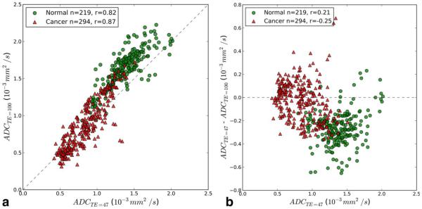
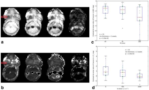
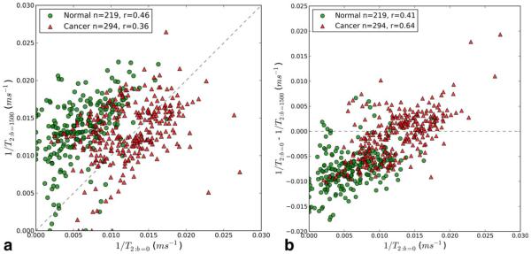
In normal prostate ROIs, the ADC at TE of 47 ms is significantly smaller than ADC at TE of 100 ms (P = 0.0003) and T2 at b-value of 0 s/mm(2) is significantly longer than T2 at b-value of 1500 s/mm(2) (P = 0.001). In cancer ROIs, average ADC and T2 values do not change as a function of TE and b-value, respectively. However, in many cancer pixels, there are large decreases in the ADC as a function of TE and large increases in T2 as a function of b-value. Cancers are more conspicuous in ADC maps at longer TEs.
Parameters derived from hybrid imaging that depend on coupled/associated values of ADC and T2 may improve the accuracy of MRI in diagnosing prostate cancer.
采用二维磁共振成像采样(即“混合多维成像”),分别研究正常前列腺组织和前列腺癌中表观扩散系数(ADC)及T2值与回波时间(TE)和b值的相关性。
本研究纳入10例经活检证实为前列腺癌的患者,均接受了3特斯拉前列腺磁共振成像检查。在b值分别为0、750和1500 s/mm²时采集扩散加权磁共振成像(DWI)数据。对于每个b值,在TE为47、75和100 ms时采集数据。分别在15个癌灶和10个正常感兴趣区(ROI)测量ADC和T2值随b值和TE的变化。采用Friedman检验来检验ADC随TE变化以及T2随b值变化的显著性。
在正常前列腺ROI中,TE为47 ms时的ADC显著小于TE为100 ms时的ADC(P = 0.0003),且b值为0 s/mm²时的T2显著长于b值为1500 s/mm²时的T2(P = 0.001)。在癌灶ROI中,平均ADC和T2值分别不随TE和b值变化。然而,在许多癌灶像素中,ADC随TE有大幅下降,T2随b值有大幅上升。在较长TE的ADC图中,癌灶更明显。
源自混合成像且依赖于ADC和T2耦合/关联值的参数可能会提高磁共振成像诊断前列腺癌的准确性。