Department of Neurological Surgery, Washington University School of Medicine , St. Louis, MO , USA.
Front Hum Neurosci. 2013 Jul 31;7:431. doi: 10.3389/fnhum.2013.00431. eCollection 2013.
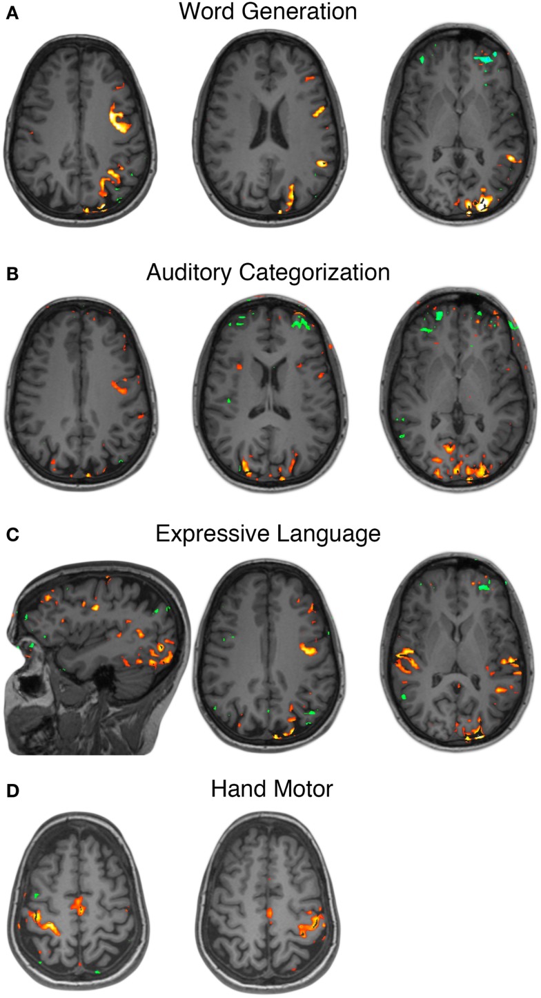
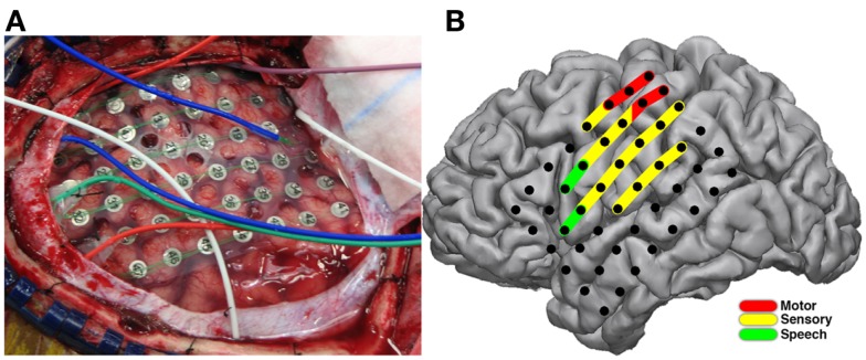
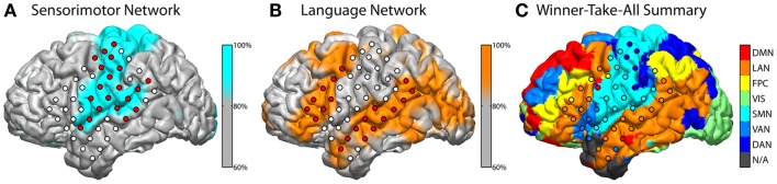
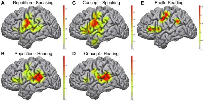
Recent advances in basic neuroscience research across a wide range of methodologies have contributed significantly to our understanding of human cortical electrophysiology and functional brain imaging. Translation of this research into clinical neurosurgery has opened doors for advanced mapping of functionality that previously was prohibitively difficult, if not impossible. Here we present the case of a unique individual with congenital blindness and medically refractory epilepsy who underwent neurosurgical treatment of her seizures. Pre-operative evaluation presented the challenge of accurately and robustly mapping the cerebral cortex for an individual with a high probability of significant cortical re-organization. Additionally, a blind individual has unique priorities in one's ability to read Braille by touch and sense the environment primarily by sound than the non-vision impaired person. For these reasons we employed additional measures to map sensory, motor, speech, language, and auditory perception by employing a number of cortical electrophysiologic mapping and functional magnetic resonance imaging methods. Our data show promising results in the application of these adjunctive methods in the pre-operative mapping of otherwise difficult to localize, and highly variable, functional cortical areas.
近年来,基础神经科学研究在各种方法学上的进展,极大地促进了我们对人类皮质电生理学和功能脑成像的理解。将这一研究转化为临床神经外科,为功能的高级定位开辟了道路,而这些功能以前是极其困难的,如果不是不可能的话。在这里,我们介绍了一个独特的个体案例,他天生失明,患有药物难治性癫痫,接受了神经外科治疗。术前评估提出了一个挑战,即如何准确而稳健地为一个极有可能发生显著皮质重组的个体绘制大脑皮质图。此外,盲人在通过触摸阅读盲文和主要通过声音感知环境方面具有独特的优先级,而不是非视觉障碍者。出于这些原因,我们采用了其他一些措施,通过使用一些皮质电生理图谱和功能磁共振成像方法来绘制感觉、运动、言语、语言和听觉感知。我们的数据显示,这些辅助方法在术前对其他难以定位和高度可变的功能皮质区域进行定位方面具有应用前景。