• 文献检索
  • 文档翻译
  • 深度研究
  • 学术资讯
  • Suppr Zotero 插件Zotero 插件
  • 邀请有礼
  • 套餐&价格
  • 历史记录
应用&插件
Suppr Zotero 插件Zotero 插件浏览器插件Mac 客户端Windows 客户端微信小程序
定价
高级版会员购买积分包购买API积分包
服务
文献检索文档翻译深度研究API 文档MCP 服务
关于我们
关于 Suppr公司介绍联系我们用户协议隐私条款
关注我们

Suppr 超能文献

核心技术专利:CN118964589B侵权必究
粤ICP备2023148730 号-1Suppr @ 2026

文献检索

告别复杂PubMed语法,用中文像聊天一样搜索,搜遍4000万医学文献。AI智能推荐,让科研检索更轻松。

立即免费搜索

文件翻译

保留排版,准确专业,支持PDF/Word/PPT等文件格式,支持 12+语言互译。

免费翻译文档

深度研究

AI帮你快速写综述,25分钟生成高质量综述,智能提取关键信息,辅助科研写作。

立即免费体验

癫痫手术后复发的复杂临床病例的术前 EEG-fMRI

Presurgical EEG-fMRI in a complex clinical case with seizure recurrence after epilepsy surgery.

机构信息

School of Biomedical Engineering, Capital Medical University, Beijing, People's Republic of China.

出版信息

Neuropsychiatr Dis Treat. 2013;9:1003-10. doi: 10.2147/NDT.S47099. Epub 2013 Jul 26.

DOI:10.2147/NDT.S47099
PMID:23926432
原文链接:https://pmc.ncbi.nlm.nih.gov/articles/PMC3732198/
Abstract

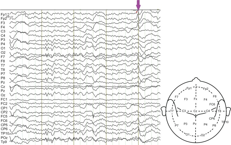
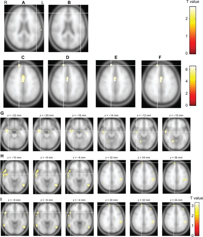
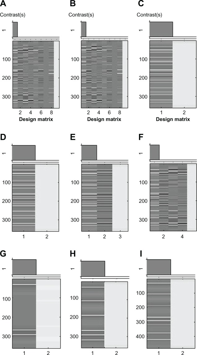
Epilepsy surgery has improved over the last decade, but non-seizure-free outcome remains at 10%-40% in temporal lobe epilepsy (TLE) and 40%-60% in extratemporal lobe epilepsy (ETLE). This paper reports a complex multifocal case. With a normal magnetic resonance imaging (MRI) result and nonlocalizing electroencephalography (EEG) findings (bilateral TLE and ETLE, with more interictal epileptiform discharges [IEDs] in the right frontal and temporal regions), a presurgical EEG-functional MRI (fMRI) was performed before the intraoperative intracranial EEG (icEEG) monitoring (icEEG with right hemispheric coverage). Our previous EEG-fMRI analysis results (IEDs in the left hemisphere alone) were contradictory to the EEG and icEEG findings (IEDs in the right frontal and temporal regions). Thus, the EEG-fMRI data were reanalyzed with newly identified IED onsets and different fMRI model options. The reanalyzed EEG-fMRI findings were largely concordant with those of EEG and icEEG, and the failure of our previous EEG-fMRI analysis may lie in the inaccurate identification of IEDs and wrong usage of model options. The right frontal and temporal regions were resected in surgery, and dual pathology (hippocampus sclerosis and focal cortical dysplasia in the extrahippocampal region) was found. The patient became seizure-free for 3 months, but his seizures restarted after antiepileptic drugs (AEDs) were stopped. The seizures were not well controlled after resuming AEDs. Postsurgical EEGs indicated that ictal spikes in the right frontal and temporal regions reduced, while those in the left hemisphere became prominent. This case suggested that (1) EEG-fMRI is valuable in presurgical evaluation, but requires caution; and (2) the intact seizure focus in the remaining brain may cause the non-seizure-free outcome.

摘要

癫痫手术在过去十年中有所改进,但颞叶癫痫(TLE)的无发作结果仍为 10%-40%,而外侧颞叶癫痫(ETLE)的无发作结果为 40%-60%。本文报告了一个复杂的多灶性病例。患者磁共振成像(MRI)结果正常,脑电图(EEG)无定位发现(双侧 TLE 和 ETLE,右侧额颞区有更多的发作间期癫痫样放电[IEDs]),在术中颅内脑电图(icEEG)监测(右侧半球覆盖的 icEEG)前进行了术前 EEG-功能磁共振成像(fMRI)。我们之前的 EEG-fMRI 分析结果(仅左半球有 IEDs)与 EEG 和 icEEG 结果(右侧额颞区的 IEDs)相矛盾。因此,使用新确定的 IED 发作和不同的 fMRI 模型选项重新分析 EEG-fMRI 数据。重新分析的 EEG-fMRI 结果与 EEG 和 icEEG 的结果基本一致,我们之前的 EEG-fMRI 分析失败可能在于 IEDs 的不准确识别和模型选项的错误使用。手术中切除了右侧额颞区,发现存在双重病变(海马硬化和海马旁区局灶性皮质发育不良)。患者术后 3 个月无发作,但停药后癫痫发作再次发作。重新开始使用 AEDs 后,癫痫发作仍未得到很好的控制。术后 EEG 显示右侧额颞区的发作棘波减少,而左侧半球的发作棘波变得明显。该病例表明:(1)EEG-fMRI 对术前评估很有价值,但需要谨慎;(2)残留大脑中完整的癫痫灶可能导致无发作结果。

https://cdn.ncbi.nlm.nih.gov/pmc/blobs/6898/3732198/3a59bfa9b951/ndt-9-1003f3.jpg
https://cdn.ncbi.nlm.nih.gov/pmc/blobs/6898/3732198/2947b342904b/ndt-9-1003f1.jpg
https://cdn.ncbi.nlm.nih.gov/pmc/blobs/6898/3732198/d86d7a4f5e5b/ndt-9-1003f2.jpg
https://cdn.ncbi.nlm.nih.gov/pmc/blobs/6898/3732198/3a59bfa9b951/ndt-9-1003f3.jpg
https://cdn.ncbi.nlm.nih.gov/pmc/blobs/6898/3732198/2947b342904b/ndt-9-1003f1.jpg
https://cdn.ncbi.nlm.nih.gov/pmc/blobs/6898/3732198/d86d7a4f5e5b/ndt-9-1003f2.jpg
https://cdn.ncbi.nlm.nih.gov/pmc/blobs/6898/3732198/3a59bfa9b951/ndt-9-1003f3.jpg

相似文献

1
Presurgical EEG-fMRI in a complex clinical case with seizure recurrence after epilepsy surgery.癫痫手术后复发的复杂临床病例的术前 EEG-fMRI
Neuropsychiatr Dis Treat. 2013;9:1003-10. doi: 10.2147/NDT.S47099. Epub 2013 Jul 26.
2
Epileptic networks in focal cortical dysplasia revealed using electroencephalography-functional magnetic resonance imaging.利用脑电图-功能磁共振成像揭示局灶性皮质发育不良中的癫痫网络。
Ann Neurol. 2011 Nov;70(5):822-37. doi: 10.1002/ana.22535.
3
Mapping Epileptic Networks Using Simultaneous Intracranial EEG-fMRI.使用同步颅内脑电图-功能磁共振成像绘制癫痫网络
Front Neurol. 2021 Sep 21;12:693504. doi: 10.3389/fneur.2021.693504. eCollection 2021.
4
Resecting critical nodes from an epileptogenic circuit in refractory focal-onset epilepsy patients using subtraction ictal SPECT coregistered to MRI.在耐药性局灶性发作性癫痫患者中,使用减去发作期 SPECT 与 MRI 配准的方法切除致痫回路中的关键节点。
J Neurosurg. 2016 Dec;125(6):1565-1576. doi: 10.3171/2015.6.JNS141719. Epub 2016 Mar 18.
5
An analysis of clinical seizure patterns and their localizing value in frontal and temporal lobe epilepsies.额叶和颞叶癫痫的临床发作模式及其定位价值分析
Brain. 1996 Feb;119 ( Pt 1):17-40. doi: 10.1093/brain/119.1.17.
6
Co-localization between the BOLD response and epileptiform discharges recorded by simultaneous intracranial EEG-fMRI at 3 T.3T 下同步颅内脑电图-功能磁共振成像记录的血氧水平依赖(BOLD)反应与癫痫样放电之间的共定位
Neuroimage Clin. 2015 Mar 7;7:755-63. doi: 10.1016/j.nicl.2015.03.002. eCollection 2015.
7
Identifying the affected hemisphere with a multimodal approach in MRI-positive or negative, unilateral or bilateral temporal lobe epilepsy.使用多模态方法在 MRI 阳性或阴性、单侧或双侧颞叶癫痫中识别受影响的半球。
Neuropsychiatr Dis Treat. 2014 Jan 7;10:71-81. doi: 10.2147/NDT.S56404. eCollection 2014.
8
Lateralizing value of interictal spikes on overnight sleep-EEG studies in temporal lobe epilepsy.颞叶癫痫患者夜间睡眠脑电图研究中发作间期棘波的定侧价值
Epilepsia. 1999 Nov;40(11):1587-92. doi: 10.1111/j.1528-1157.1999.tb02044.x.
9
EEG correlated functional MRI and postoperative outcome in focal epilepsy.脑电相关功能磁共振成像与局灶性癫痫的术后结果。
J Neurol Neurosurg Psychiatry. 2010 Aug;81(8):922-7. doi: 10.1136/jnnp.2009.196253. Epub 2010 Jun 14.
10
The Role of Interictal Epileptiform Discharges in Epilepsy Surgery Outcome.发作间期癫痫样放电在癫痫手术疗效中的作用
Int J Prev Med. 2019 Jun 7;10:101. doi: 10.4103/ijpvm.IJPVM_237_18. eCollection 2019.

引用本文的文献

1
Spatio-temporal independent component classification for localization of seizure onset zone.用于癫痫发作起始区定位的时空独立成分分类
Front Neurol. 2025 Jun 6;16:1515484. doi: 10.3389/fneur.2025.1515484. eCollection 2025.
2
Localization of Epileptic Foci Based on Simultaneous EEG-fMRI Data.基于同步脑电图-功能磁共振成像数据的癫痫病灶定位
Front Neurol. 2021 Apr 27;12:645594. doi: 10.3389/fneur.2021.645594. eCollection 2021.

本文引用的文献

1
Temporal lobe epilepsy surgery failures: a review.颞叶癫痫手术失败:综述
Epilepsy Res Treat. 2012;2012:201651. doi: 10.1155/2012/201651. Epub 2012 Apr 22.
2
EEG-fMRI validation studies in comparison with icEEG: a review.脑电-功能磁共振成像验证研究与颅内脑电图的比较:综述。
Int J Psychophysiol. 2012 Jun;84(3):233-9. doi: 10.1016/j.ijpsycho.2012.01.021. Epub 2012 Feb 15.
3
With or without spikes: localization of focal epileptic activity by simultaneous electroencephalography and functional magnetic resonance imaging.
有或无棘波:同步脑电图和功能磁共振成像定位局灶性癫痫活动。
Brain. 2011 Oct;134(Pt 10):2867-86. doi: 10.1093/brain/awr156. Epub 2011 Jul 12.
4
Advances in MRI for 'cryptogenic' epilepsies.MRI 技术在“隐源性”癫痫中的应用进展。
Nat Rev Neurol. 2011 Feb;7(2):99-108. doi: 10.1038/nrneurol.2010.199. Epub 2011 Jan 18.
5
Integrating EEG and fMRI in epilepsy.将 EEG 和 fMRI 整合到癫痫中。
Neuroimage. 2011 Feb 14;54(4):2719-31. doi: 10.1016/j.neuroimage.2010.11.038. Epub 2010 Nov 23.
6
Imaging haemodynamic changes related to seizures: comparison of EEG-based general linear model, independent component analysis of fMRI and intracranial EEG.成像与癫痫相关的血流动力学变化:基于脑电图的广义线性模型、功能磁共振成像独立成分分析和颅内脑电图的比较。
Neuroimage. 2010 Oct 15;53(1):196-205. doi: 10.1016/j.neuroimage.2010.05.064. Epub 2010 Jun 4.
7
Present and future of simultaneous EEG-fMRI.脑电与功能磁共振成像的现状与未来。
MAGMA. 2010 Dec;23(5-6):309-16. doi: 10.1007/s10334-009-0196-9. Epub 2010 Jan 26.
8
EEG-fMRI: adding to standard evaluations of patients with nonlesional frontal lobe epilepsy.脑电图-功能磁共振成像:用于补充非病变性额叶癫痫患者的标准评估。
Neurology. 2009 Dec 8;73(23):2023-30. doi: 10.1212/WNL.0b013e3181c55d17.
9
How wrong can we be? The effect of inaccurate mark-up of EEG/fMRI studies in epilepsy.我们可能会错到什么程度?脑电图/功能磁共振成像研究中癫痫标记不准确的影响。
Clin Neurophysiol. 2009 Sep;120(9):1637-47. doi: 10.1016/j.clinph.2009.04.025. Epub 2009 Jul 25.
10
Different structures involved during ictal and interictal epileptic activity in malformations of cortical development: an EEG-fMRI study.皮质发育畸形患者发作期和发作间期癫痫活动涉及的不同结构:一项脑电图-功能磁共振成像研究
Brain. 2008 Aug;131(Pt 8):2042-60. doi: 10.1093/brain/awn145. Epub 2008 Jul 16.