Department of Biomedical Engineering, Duke University, Durham, North Carolina, USA.
Ultrasound Med Biol. 2013 Oct;39(10):1861-74. doi: 10.1016/j.ultrasmedbio.2013.03.029. Epub 2013 Aug 9.
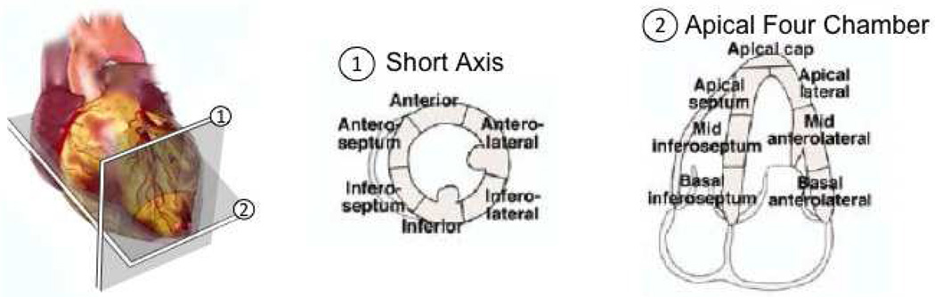
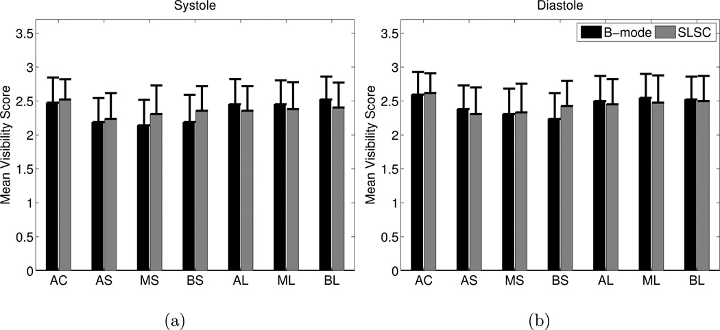
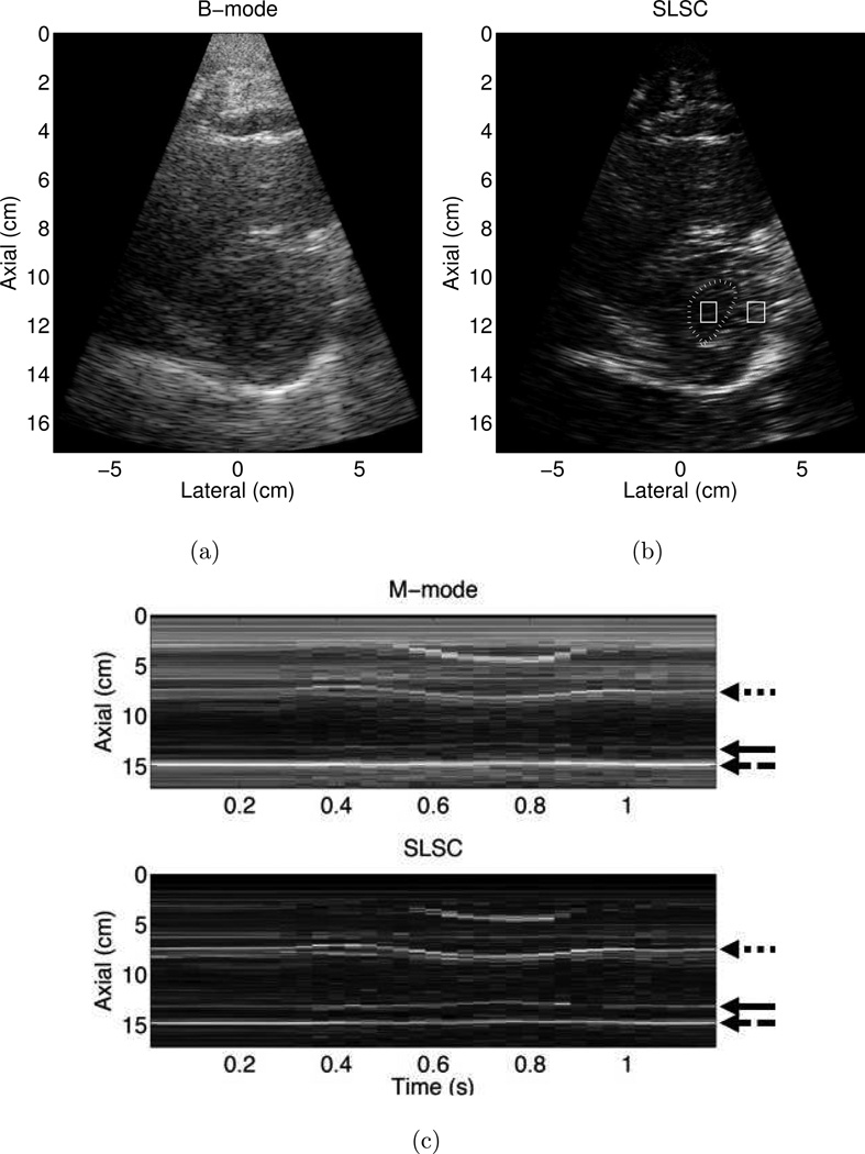
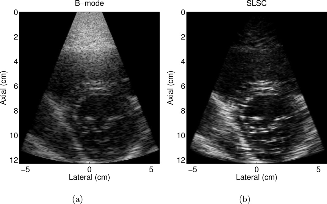
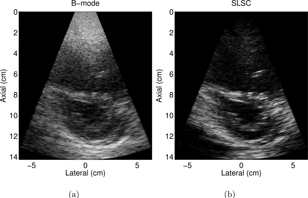
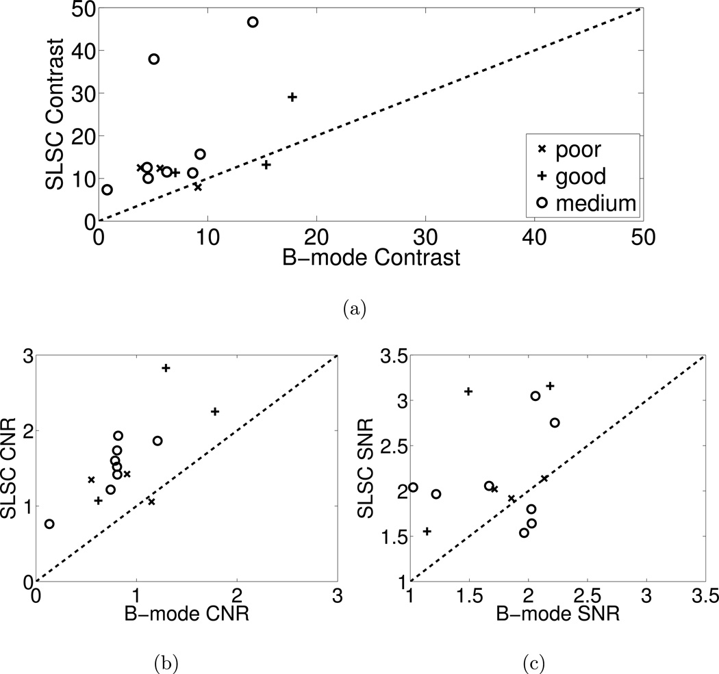
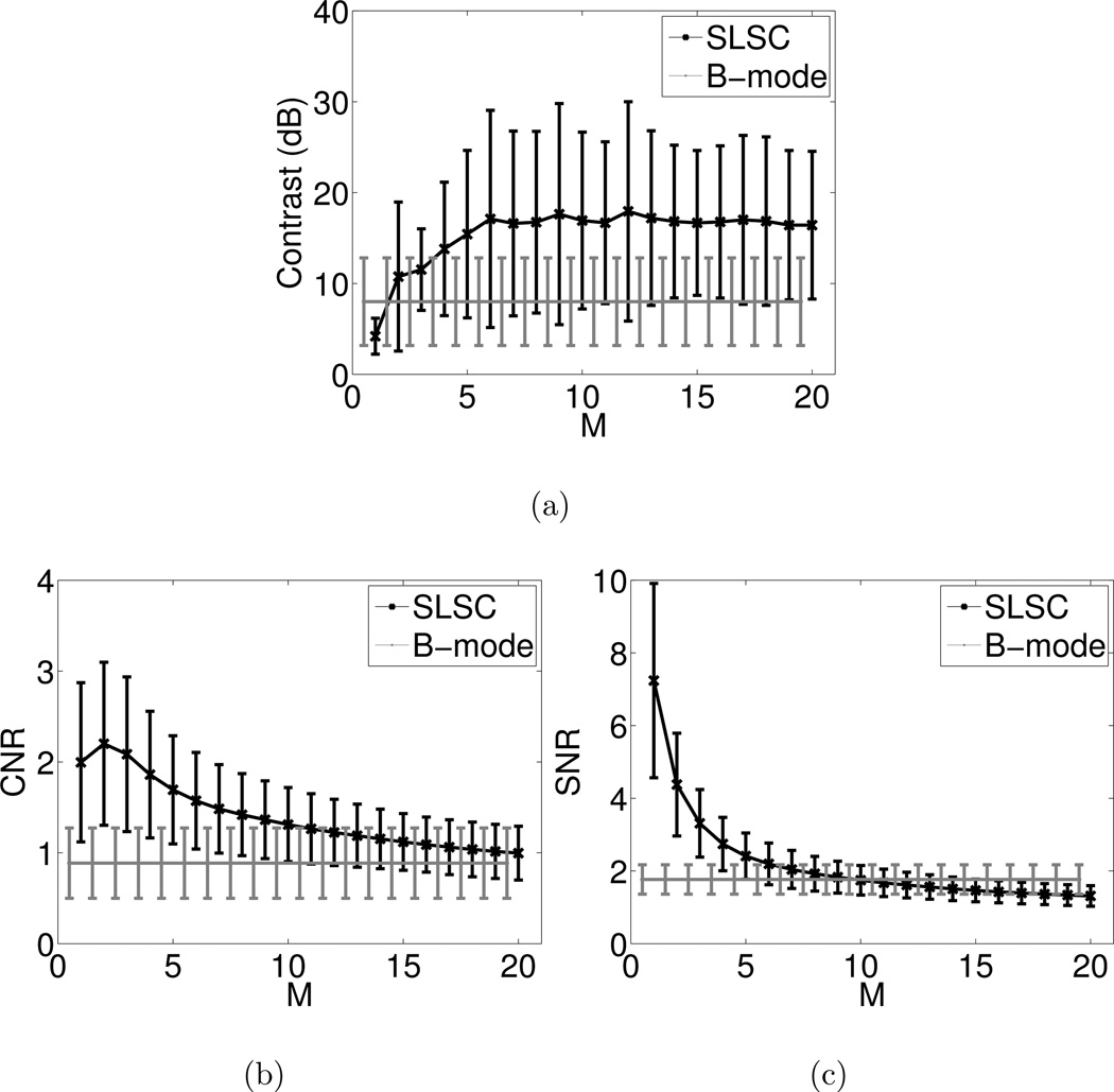
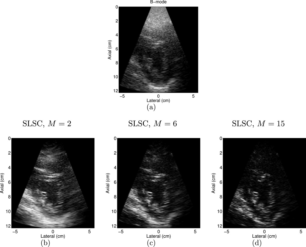
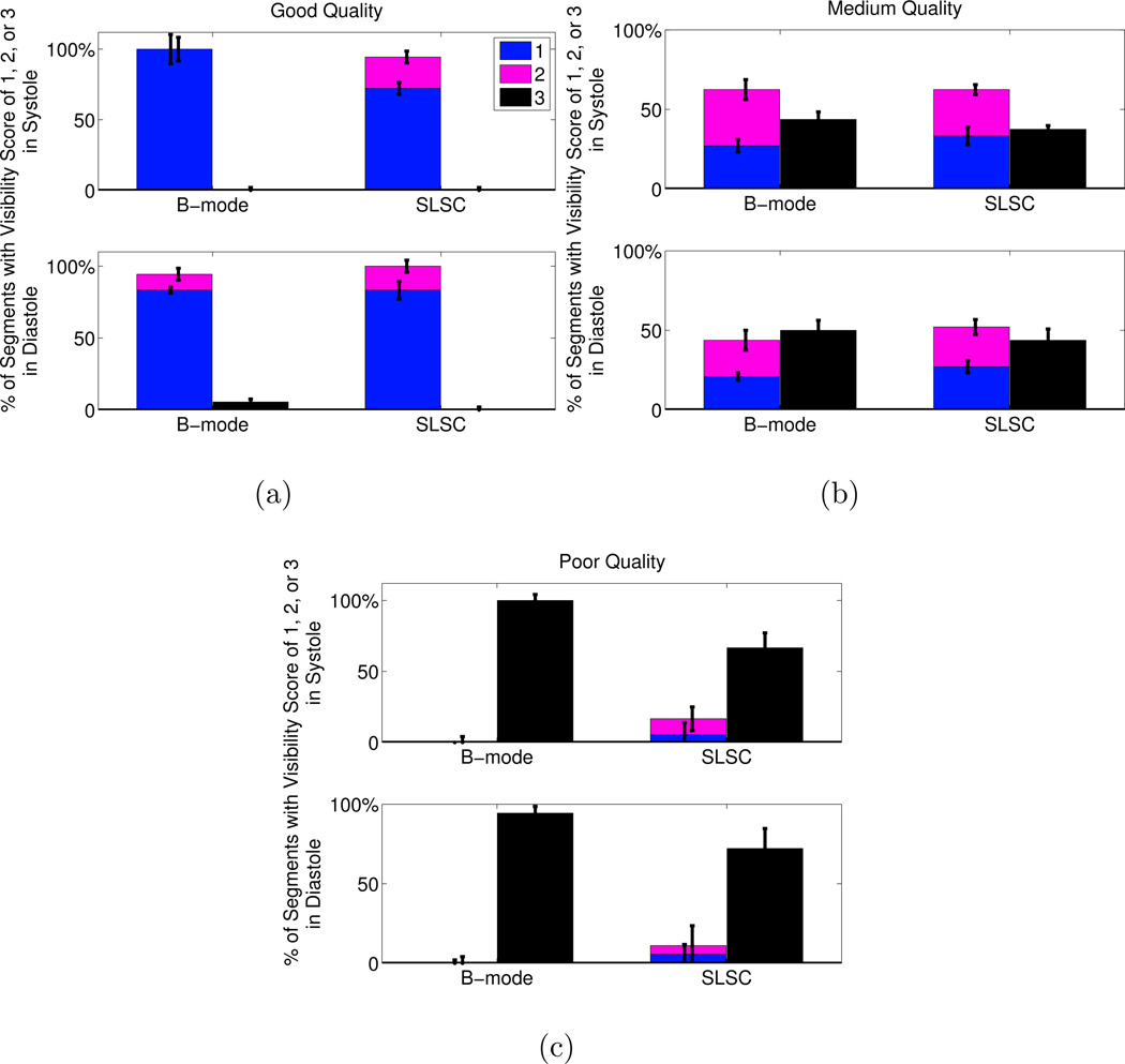
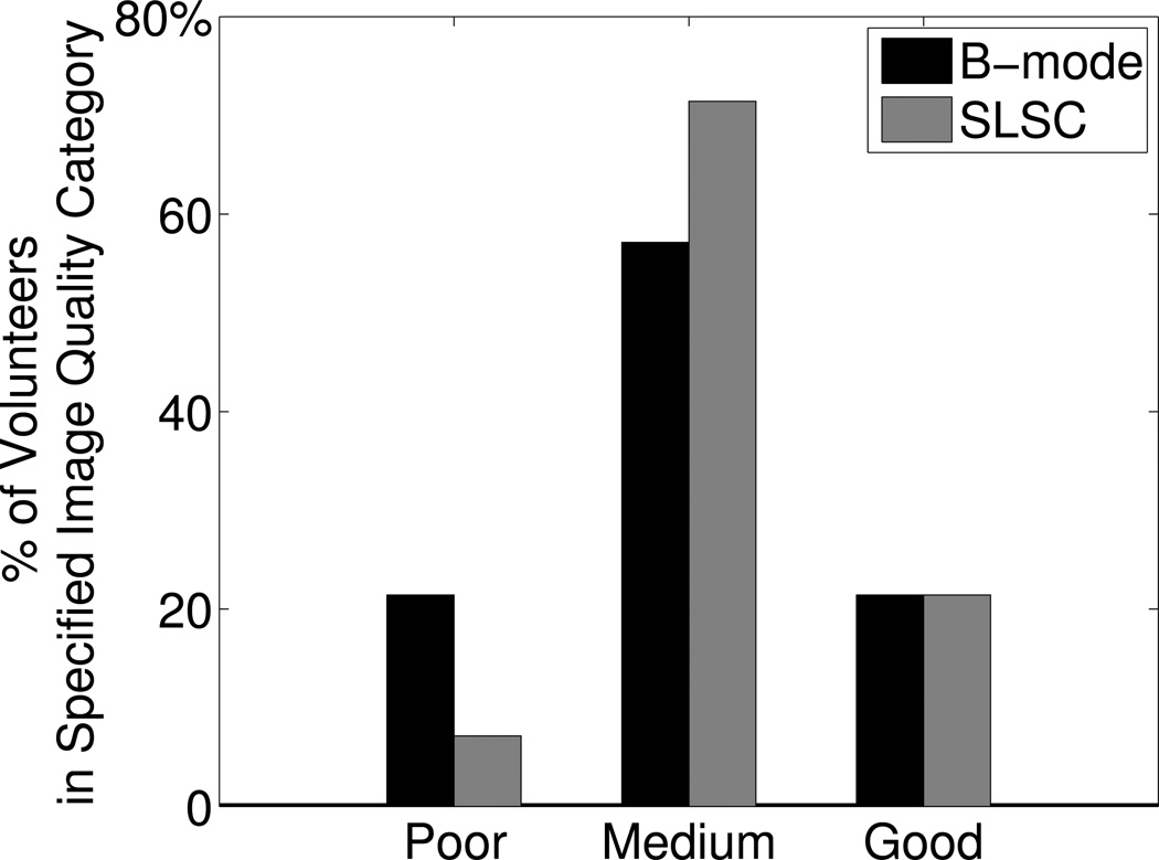
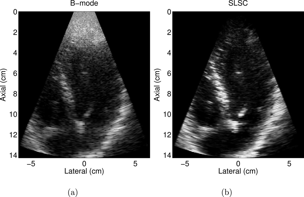
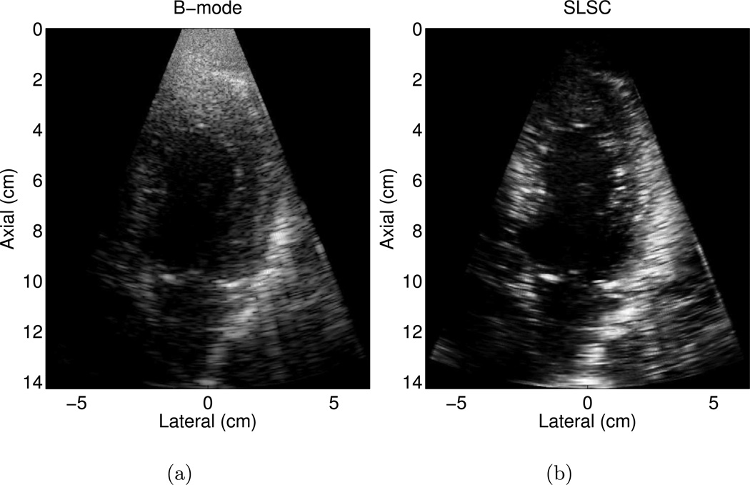
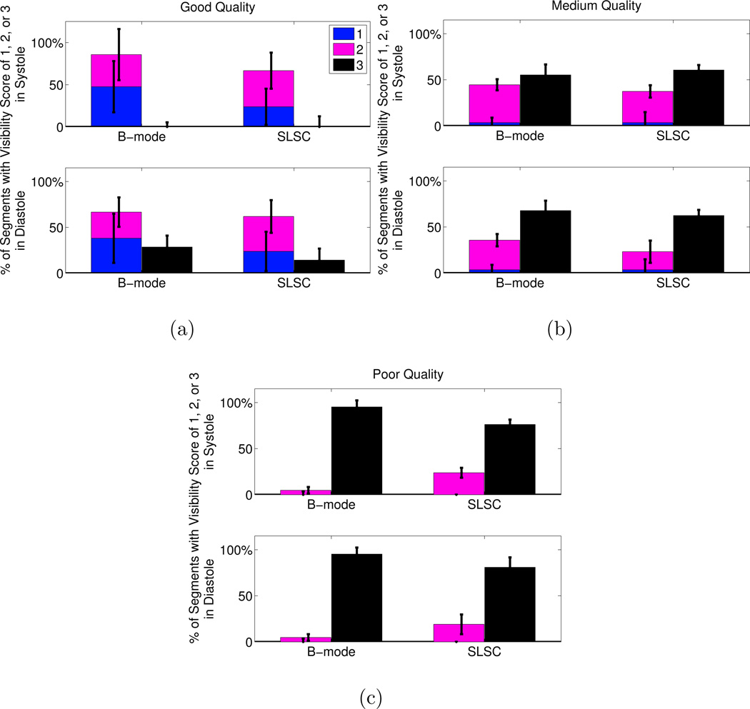
Short-lag spatial coherence (SLSC) imaging is a novel beamforming technique that reduces acoustic clutter in ultrasound images. A clinical study was conducted to investigate clutter reduction and endocardial border detection in cardiac SLSC images. Individual channel echo data were acquired from the left ventricle of 14 volunteers, after informed consent and institutional review board approval. Paired B-mode and SLSC images were created from these data. Contrast, contrast-to-noise, and signal-to-noise ratios were measured in paired images, and these metrics were improved with SLSC imaging in most cases. Three cardiology fellows rated the visibility of endocardial segments in randomly ordered B-mode and SLSC cine loops. SLSC imaging offered 22%-33% improvement (p < 0.05) in endocardial border visibility when B-mode image quality was poor (i.e., 80% or more of the endocardial segments could not be visualized by the three reviewers). The percentage of volunteers with poor-quality images was decreased from 21% to 7% with the SLSC beamformer. Results suggest that SLSC imaging has the potential to improve clinical cardiac assessments that are challenged by clutter.
短延迟空间相干(SLSC)成像是一种新颖的波束形成技术,可减少超声图像中的声散射。进行了一项临床研究,以调查心脏 SLSC 图像中的杂波减少和心内膜边界检测。在获得知情同意书和机构审查委员会批准后,从 14 名志愿者的左心室采集单个通道回波数据。从这些数据中创建了配对的 B 模式和 SLSC 图像。在配对图像中测量了对比度、对比度噪声比和信噪比,并且在大多数情况下,SLSC 成像都提高了这些指标。三名心脏病学研究员对随机顺序的 B 模式和 SLSC 电影循环中的心内膜段的可见性进行了评分。当 B 模式图像质量较差(即三位审阅者无法看到 80%或更多的心内膜段)时,SLSC 成像在心内膜边界可见性方面提供了 22%-33%的改善(p < 0.05)。使用 SLSC 波束形成器,图像质量差的志愿者百分比从 21%降至 7%。结果表明,SLSC 成像有可能改善因杂波而受到挑战的临床心脏评估。