Hodneland Erlend, Kögel Tanja, Frei Dominik Michael, Gerdes Hans-Hermann, Lundervold Arvid
Department of Biomedicine, University of Bergen, Bergen, Norway.
Source Code Biol Med. 2013 Aug 9;8(1):16. doi: 10.1186/1751-0473-8-16.
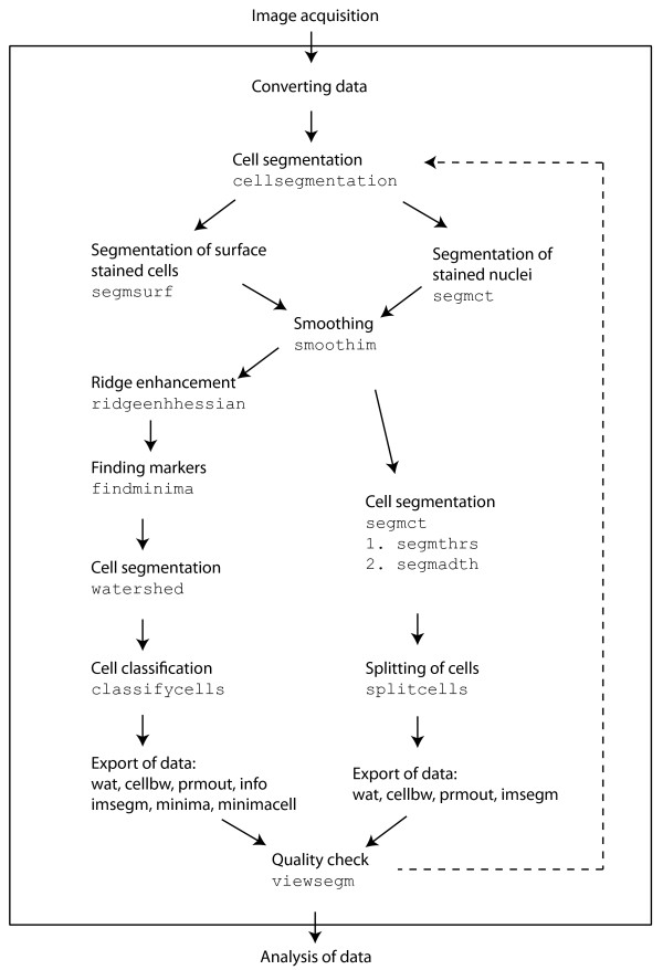
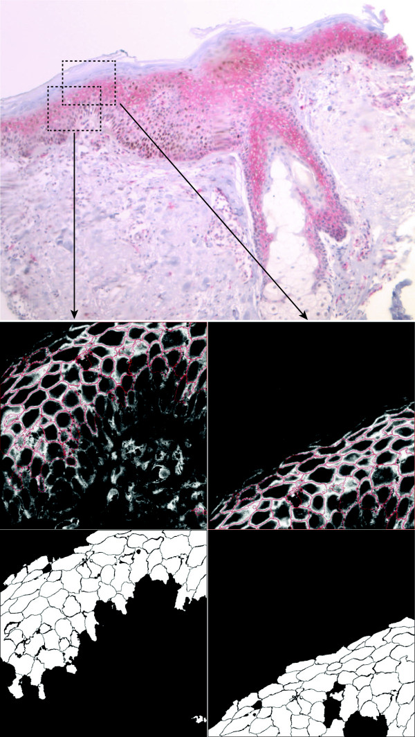
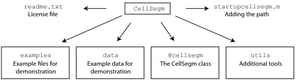
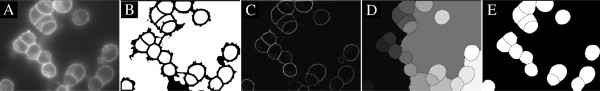
: The application of fluorescence microscopy in cell biology often generates a huge amount of imaging data. Automated whole cell segmentation of such data enables the detection and analysis of individual cells, where a manual delineation is often time consuming, or practically not feasible. Furthermore, compared to manual analysis, automation normally has a higher degree of reproducibility. CellSegm, the software presented in this work, is a Matlab based command line software toolbox providing an automated whole cell segmentation of images showing surface stained cells, acquired by fluorescence microscopy. It has options for both fully automated and semi-automated cell segmentation. Major algorithmic steps are: (i) smoothing, (ii) Hessian-based ridge enhancement, (iii) marker-controlled watershed segmentation, and (iv) feature-based classfication of cell candidates. Using a wide selection of image recordings and code snippets, we demonstrate that CellSegm has the ability to detect various types of surface stained cells in 3D. After detection and outlining of individual cells, the cell candidates can be subject to software based analysis, specified and programmed by the end-user, or they can be analyzed by other software tools. A segmentation of tissue samples with appropriate characteristics is also shown to be resolvable in CellSegm. The command-line interface of CellSegm facilitates scripting of the separate tools, all implemented in Matlab, offering a high degree of flexibility and tailored workflows for the end-user. The modularity and scripting capabilities of CellSegm enable automated workflows and quantitative analysis of microscopic data, suited for high-throughput image based screening.
荧光显微镜在细胞生物学中的应用常常会产生大量的成像数据。对这类数据进行全细胞自动分割能够实现对单个细胞的检测与分析,而手动勾勒细胞轮廓往往耗时较长,甚至在实际操作中不可行。此外,与手动分析相比,自动化分析通常具有更高的可重复性。CellSegm是本文介绍的一款软件,它是一个基于Matlab的命令行软件工具箱,可对通过荧光显微镜获取的表面染色细胞图像进行全细胞自动分割。它提供了全自动和半自动细胞分割选项。主要算法步骤包括:(i)平滑处理,(ii)基于黑塞矩阵的脊增强,(iii)标记控制的分水岭分割,以及(iv)基于特征的细胞候选物分类。通过广泛选择图像记录和代码片段,我们证明CellSegm能够在三维空间中检测各种类型的表面染色细胞。在检测并勾勒出单个细胞后,细胞候选物可以由终端用户指定并编程进行基于软件的分析,也可以由其他软件工具进行分析。CellSegm还能够对具有适当特征的组织样本进行分割。CellSegm的命令行界面便于对所有用Matlab实现的单独工具进行脚本编写,为终端用户提供了高度的灵活性和定制化工作流程。CellSegm的模块化和脚本编写功能能够实现自动化工作流程以及对微观数据的定量分析,适用于基于高通量图像的筛选。