• 文献检索
  • 文档翻译
  • 深度研究
  • 学术资讯
  • Suppr Zotero 插件Zotero 插件
  • 邀请有礼
  • 套餐&价格
  • 历史记录
应用&插件
Suppr Zotero 插件Zotero 插件浏览器插件Mac 客户端Windows 客户端微信小程序
定价
高级版会员购买积分包购买API积分包
服务
文献检索文档翻译深度研究API 文档MCP 服务
关于我们
关于 Suppr公司介绍联系我们用户协议隐私条款
关注我们

Suppr 超能文献

核心技术专利:CN118964589B侵权必究
粤ICP备2023148730 号-1Suppr @ 2026

文献检索

告别复杂PubMed语法,用中文像聊天一样搜索,搜遍4000万医学文献。AI智能推荐,让科研检索更轻松。

立即免费搜索

文件翻译

保留排版,准确专业,支持PDF/Word/PPT等文件格式,支持 12+语言互译。

免费翻译文档

深度研究

AI帮你快速写综述,25分钟生成高质量综述,智能提取关键信息,辅助科研写作。

立即免费体验

一种用于半自动基于细胞的对象共定位分析的通用工具箱。

A versatile toolbox for semi-automatic cell-by-cell object-based colocalization analysis.

机构信息

Laboratory of Neural Development and Optical Recording (NDEVOR), Division of Physiology, Department of Molecular Medicine, University of Oslo, Blindern, 1105, Oslo, Norway.

出版信息

Sci Rep. 2020 Nov 4;10(1):19027. doi: 10.1038/s41598-020-75835-7.

DOI:10.1038/s41598-020-75835-7
PMID:33149236
原文链接:https://pmc.ncbi.nlm.nih.gov/articles/PMC7643144/
Abstract

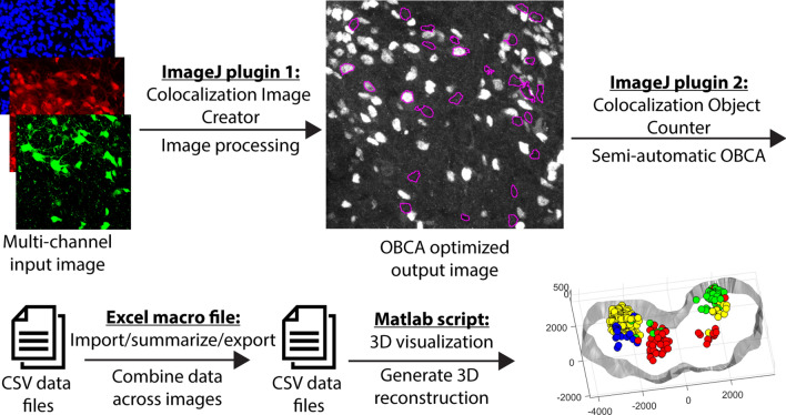
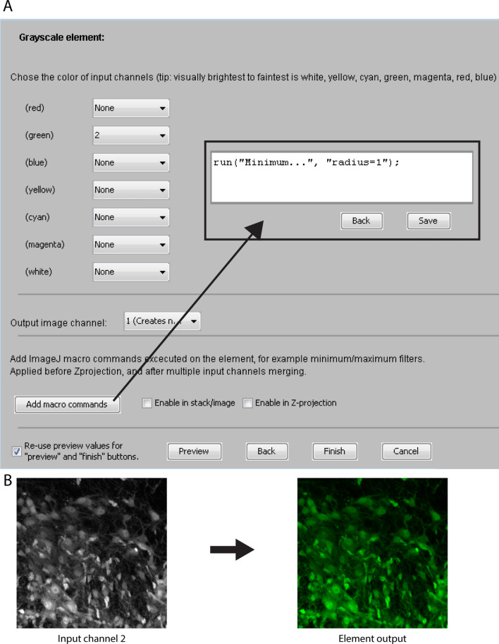
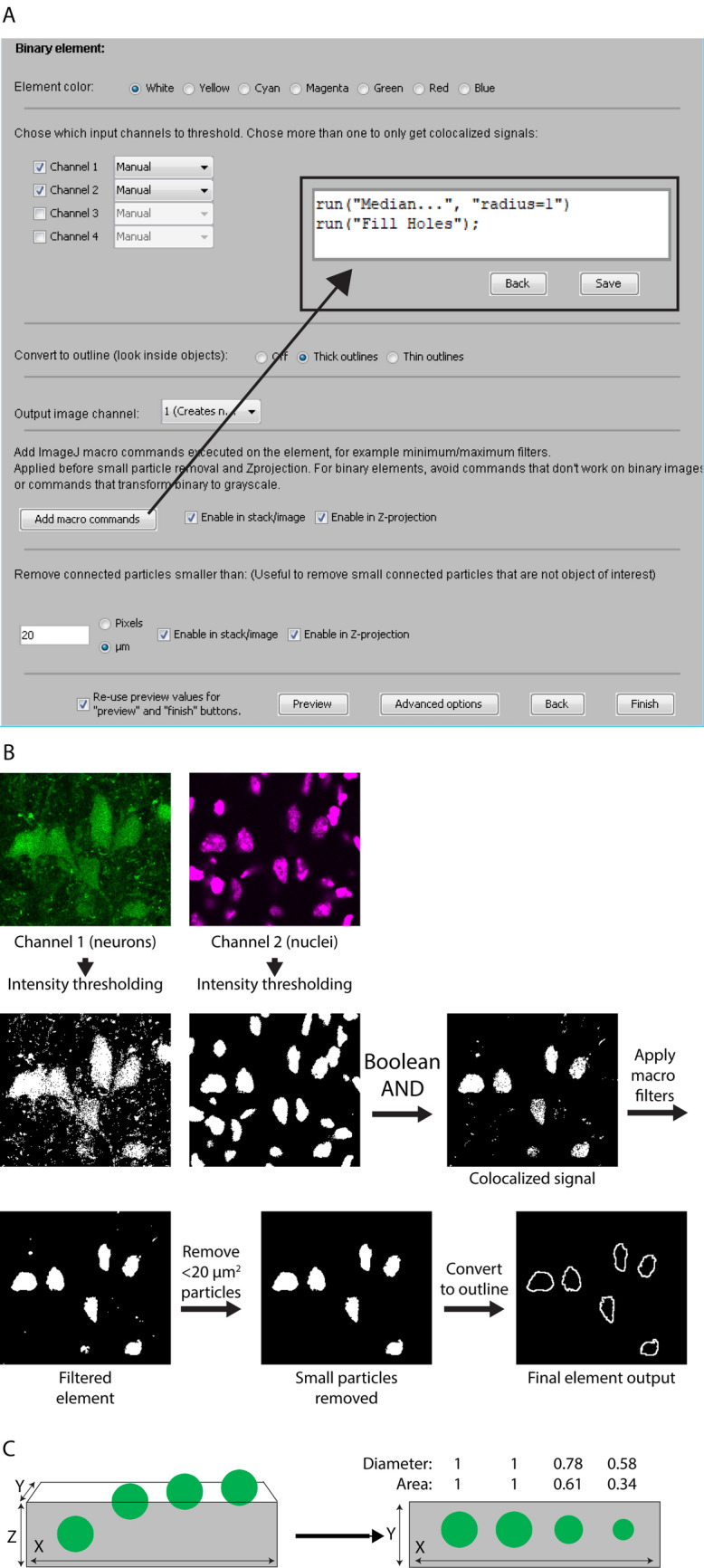
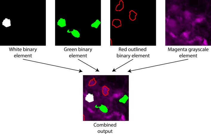
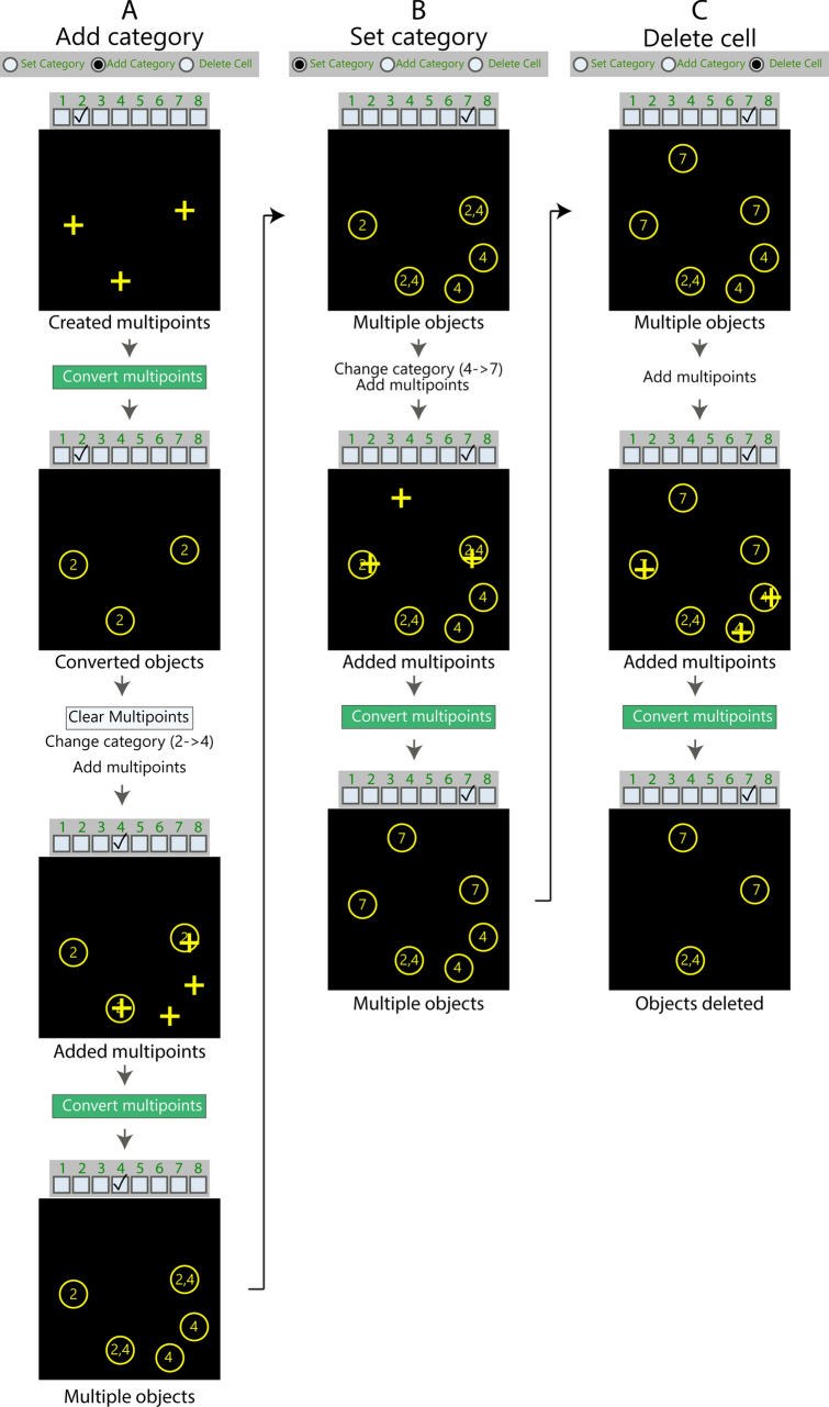
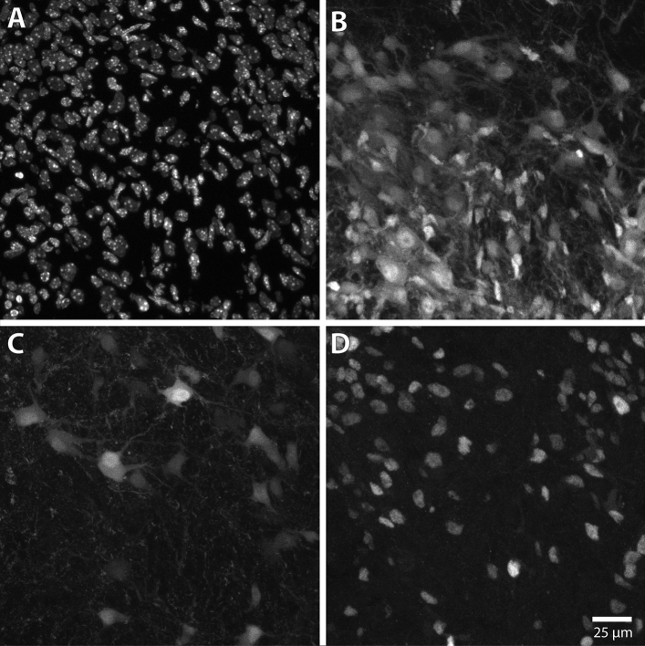
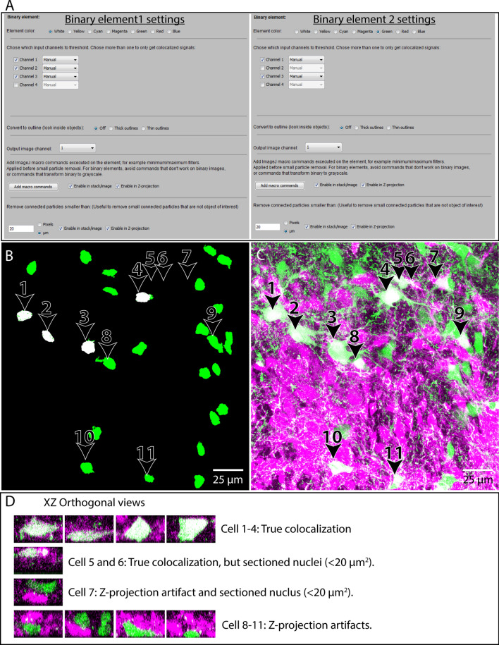
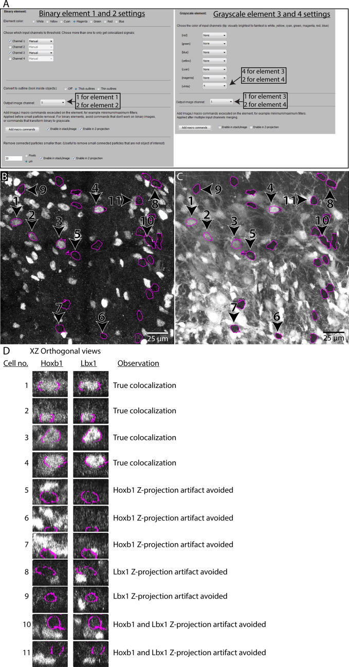
Differential fluorescence labeling and multi-fluorescence imaging followed by colocalization analysis is commonly used to investigate cellular heterogeneity in situ. This is particularly important when investigating the biology of tissues with diverse cell types. Object-based colocalization analysis (OBCA) tools can employ automatic approaches, which are sensitive to errors in cell segmentation, or manual approaches, which can be impractical and tedious. Here, we present a novel set of tools for OBCA using a semi-automatic approach, consisting of two ImageJ plugins, a Microsoft Excel macro, and a MATLAB script. One ImageJ plugin enables customizable processing of multichannel 3D images for enhanced visualization of features relevant to OBCA, and another enables semi-automatic colocalization quantification. The Excel macro and the MATLAB script enable data organization and 3D visualization of object data across image series. The tools are well suited for experiments involving complex and large image data sets, and can be used in combination or as individual components, allowing flexible, efficient and accurate OBCA. Here we demonstrate their utility in immunohistochemical analyses of the developing central nervous system, which is characterized by complexity in the number and distribution of cell types, and by high cell packing densities, which can both create challenging situations for OBCA.

摘要

差异荧光标记和多荧光成像后进行共定位分析通常用于原位研究细胞异质性。当研究具有多种细胞类型的组织生物学时,这一点尤为重要。基于对象的共定位分析(OBCA)工具可以采用自动方法,这些方法容易受到细胞分割错误的影响,也可以采用手动方法,但手动方法不切实际且繁琐。在这里,我们提出了一种使用半自动方法的新型 OBCA 工具集,包括两个 ImageJ 插件、一个 Microsoft Excel 宏和一个 MATLAB 脚本。一个 ImageJ 插件可用于对多通道 3D 图像进行可定制的处理,以增强与 OBCA 相关的特征的可视化效果,另一个插件则可用于半自动共定位定量。Excel 宏和 MATLAB 脚本可用于在图像系列之间组织和可视化对象数据。这些工具非常适合涉及复杂和大型图像数据集的实验,可以组合使用或作为单独的组件,从而实现灵活、高效和准确的 OBCA。在这里,我们展示了它们在发育中的中枢神经系统免疫组织化学分析中的应用,该系统的特点是细胞类型的数量和分布复杂,细胞密度高,这两者都会给 OBCA 带来挑战。

https://cdn.ncbi.nlm.nih.gov/pmc/blobs/3656/7643144/378685dbe7eb/41598_2020_75835_Fig12_HTML.jpg
https://cdn.ncbi.nlm.nih.gov/pmc/blobs/3656/7643144/cea00fe53ef4/41598_2020_75835_Fig1_HTML.jpg
https://cdn.ncbi.nlm.nih.gov/pmc/blobs/3656/7643144/169f6a6ee4c2/41598_2020_75835_Fig2_HTML.jpg
https://cdn.ncbi.nlm.nih.gov/pmc/blobs/3656/7643144/88115dcf5860/41598_2020_75835_Fig3_HTML.jpg
https://cdn.ncbi.nlm.nih.gov/pmc/blobs/3656/7643144/4546d4f07305/41598_2020_75835_Fig4_HTML.jpg
https://cdn.ncbi.nlm.nih.gov/pmc/blobs/3656/7643144/d28b80323e29/41598_2020_75835_Fig5_HTML.jpg
https://cdn.ncbi.nlm.nih.gov/pmc/blobs/3656/7643144/4b7250ebe734/41598_2020_75835_Fig6_HTML.jpg
https://cdn.ncbi.nlm.nih.gov/pmc/blobs/3656/7643144/6afbeefa09b3/41598_2020_75835_Fig7_HTML.jpg
https://cdn.ncbi.nlm.nih.gov/pmc/blobs/3656/7643144/2624ab79beb4/41598_2020_75835_Fig8_HTML.jpg
https://cdn.ncbi.nlm.nih.gov/pmc/blobs/3656/7643144/19be781af329/41598_2020_75835_Fig9_HTML.jpg
https://cdn.ncbi.nlm.nih.gov/pmc/blobs/3656/7643144/d6ffc0d7c1e0/41598_2020_75835_Fig10_HTML.jpg
https://cdn.ncbi.nlm.nih.gov/pmc/blobs/3656/7643144/0a33ccc3a93a/41598_2020_75835_Fig11_HTML.jpg
https://cdn.ncbi.nlm.nih.gov/pmc/blobs/3656/7643144/378685dbe7eb/41598_2020_75835_Fig12_HTML.jpg

相似文献

1
A versatile toolbox for semi-automatic cell-by-cell object-based colocalization analysis.一种用于半自动基于细胞的对象共定位分析的通用工具箱。
Sci Rep. 2020 Nov 4;10(1):19027. doi: 10.1038/s41598-020-75835-7.
2
Slide Set: Reproducible image analysis and batch processing with ImageJ.幻灯片集:使用ImageJ进行可重复的图像分析和批处理。
Biotechniques. 2015 Nov 1;59(5):269-78. doi: 10.2144/000114351. eCollection 2015 Nov.
3
PaCeQuant: A Tool for High-Throughput Quantification of Pavement Cell Shape Characteristics.PaCeQuant:一种用于高通量量化路面细胞形状特征的工具。
Plant Physiol. 2017 Nov;175(3):998-1017. doi: 10.1104/pp.17.00961. Epub 2017 Sep 20.
4
FocAn: automated 3D analysis of DNA repair foci in image stacks acquired by confocal fluorescence microscopy.FocAn:通过共聚焦荧光显微镜获取的图像堆栈中 DNA 修复焦点的自动 3D 分析。
BMC Bioinformatics. 2020 Jan 28;21(1):27. doi: 10.1186/s12859-020-3370-8.
5
ITK-SNAP: An interactive tool for semi-automatic segmentation of multi-modality biomedical images.ITK-SNAP:一种用于多模态生物医学图像半自动分割的交互式工具。
Annu Int Conf IEEE Eng Med Biol Soc. 2016 Aug;2016:3342-3345. doi: 10.1109/EMBC.2016.7591443.
6
SlideJ: An ImageJ plugin for automated processing of whole slide images.SlideJ:一款用于自动处理全玻片图像的ImageJ插件。
PLoS One. 2017 Jul 6;12(7):e0180540. doi: 10.1371/journal.pone.0180540. eCollection 2017.
7
Comparison of two automatic cell-counting solutions for fluorescent microscopic images.两种用于荧光显微镜图像的自动细胞计数解决方案的比较。
J Microsc. 2015 Oct;260(1):107-16. doi: 10.1111/jmi.12272. Epub 2015 Jun 22.
8
Colocalization by cross-correlation, a new method of colocalization suited for super-resolution microscopy.通过互相关实现共定位,一种适用于超分辨率显微镜的共定位新方法。
BMC Bioinformatics. 2024 Feb 2;25(1):55. doi: 10.1186/s12859-024-05675-z.
9
A versatile macro-based neurohistological image analysis suite for ImageJ focused on automated and standardized user interaction and reproducible data output.一个适用于ImageJ的多功能基于宏的神经组织学图像分析套件,专注于自动化和标准化的用户交互以及可重复的数据输出。
J Neurosci Methods. 2019 Aug 1;324:108286. doi: 10.1016/j.jneumeth.2019.04.009. Epub 2019 May 4.
10
Automatic thoracic anatomy segmentation on CT images using hierarchical fuzzy models and registration.使用分层模糊模型和配准技术对CT图像进行胸部解剖结构自动分割
Med Phys. 2016 Mar;43(3):1487-500. doi: 10.1118/1.4942486.

引用本文的文献

1
The E3 ligase MEX3B forms a tripartite complex with and to determine the proliferative capacity of neural progenitor cells.E3泛素连接酶MEX3B与……形成三方复合物,以决定神经祖细胞的增殖能力。 (注:原文中“with and ”部分缺失具体内容)
Open Biol. 2025 Sep;15(9):250164. doi: 10.1098/rsob.250164. Epub 2025 Sep 10.
2
Bacterial Dynamics in Newly Settled Acropora kenti: Insights From Inoculations With Individual Probiotic Candidates.新定居的肯氏鹿角珊瑚中的细菌动态:来自单个益生菌候选菌株接种的见解
Environ Microbiol. 2025 Jul;27(7):e70143. doi: 10.1111/1462-2920.70143.
3
Massively parallel reporter assay for mapping gene-specific regulatory regions at single nucleotide resolution.

本文引用的文献

1
Molecular Profiling Defines Evolutionarily Conserved Transcription Factor Signatures of Major Vestibulospinal Neuron Groups.分子分析定义了主要前庭脊髓神经元群进化保守的转录因子特征。
eNeuro. 2019 Feb 27;6(1). doi: 10.1523/ENEURO.0475-18.2019. eCollection 2019 Jan-Feb.
2
Shared and distinct transcriptomic cell types across neocortical areas.不同脑区共有的和独特的转录组细胞类型。
Nature. 2018 Nov;563(7729):72-78. doi: 10.1038/s41586-018-0654-5. Epub 2018 Oct 31.
3
EzColocalization: An ImageJ plugin for visualizing and measuring colocalization in cells and organisms.
用于在单核苷酸分辨率下绘制基因特异性调控区域的大规模平行报告基因检测法。
bioRxiv. 2025 May 14:2025.05.13.653746. doi: 10.1101/2025.05.13.653746.
4
Discrete photoentrainment of mammalian central clock is regulated by bi-stable dynamic network in the suprachiasmatic nucleus.哺乳动物中枢生物钟的离散光同步受视交叉上核中的双稳态动态网络调控。
Nat Commun. 2025 Apr 8;16(1):3331. doi: 10.1038/s41467-025-58661-1.
5
Optimizing Colocalized Cell Counting Using Automated and Semiautomated Methods.使用自动化和半自动化方法优化共定位细胞计数
Neuroinformatics. 2025 Mar 21;23(2):25. doi: 10.1007/s12021-025-09723-8.
6
Synaptic protein expression in bipolar disorder patient-derived neurons implicates PSD-95 as a marker of lithium response.双相情感障碍患者来源神经元中的突触蛋白表达表明PSD-95是锂反应的一个标志物。
Neuropharmacology. 2025 May 1;268:110313. doi: 10.1016/j.neuropharm.2025.110313. Epub 2025 Jan 15.
7
The circular RNA circNFIX regulates MEF2C expression in muscle satellite cells in spastic cerebral palsy.环状RNA circNFIX在痉挛性脑瘫的肌肉卫星细胞中调节MEF2C的表达。
J Biol Chem. 2024 Dec;300(12):107987. doi: 10.1016/j.jbc.2024.107987. Epub 2024 Nov 13.
8
γδ T cell-mediated activation of cDC1 orchestrates CD4 Th1 cell priming in malaria.γδ T 细胞介导的 cDC1 激活在疟疾中协调 CD4 Th1 细胞的启动。
Front Immunol. 2024 Aug 15;15:1426316. doi: 10.3389/fimmu.2024.1426316. eCollection 2024.
9
Elucidating the mechano-molecular dynamics of TRAP activity using CRISPR/Cas9 mediated fluorescent reporter mice.利用CRISPR/Cas9介导的荧光报告基因小鼠阐明TRAP活性的机械分子动力学。
Heliyon. 2024 Jun 13;10(12):e32949. doi: 10.1016/j.heliyon.2024.e32949. eCollection 2024 Jun 30.
10
Y chromosome shredding in Anopheles gambiae: Insight into the cellular dynamics of a novel synthetic sex ratio distorter.Y 染色体碎裂在冈比亚按蚊中的作用:新型合成性别比例干扰因子的细胞动态学研究。
PLoS Genet. 2024 Jun 7;20(6):e1011303. doi: 10.1371/journal.pgen.1011303. eCollection 2024 Jun.
EzColocalization:一款用于可视化和测量细胞和生物体内共定位的 ImageJ 插件。
Sci Rep. 2018 Oct 25;8(1):15764. doi: 10.1038/s41598-018-33592-8.
4
Image co-localization - co-occurrence versus correlation.图像共定位——共现与相关。
J Cell Sci. 2018 Feb 8;131(3):jcs211847. doi: 10.1242/jcs.211847.
5
QuPath: Open source software for digital pathology image analysis.QuPath:用于数字病理学图像分析的开源软件。
Sci Rep. 2017 Dec 4;7(1):16878. doi: 10.1038/s41598-017-17204-5.
6
MatCol: a tool to measure fluorescence signal colocalisation in biological systems.MatCol:一种用于测量生物系统中荧光信号共定位的工具。
Sci Rep. 2017 Aug 21;7(1):8879. doi: 10.1038/s41598-017-08786-1.
7
Neuroscience in the third dimension: shedding new light on the brain with tissue clearing.三维空间中的神经科学:组织透明化技术为大脑研究带来新曙光
Mol Brain. 2017 Jul 20;10(1):33. doi: 10.1186/s13041-017-0314-y.
8
Quantitating the cell: turning images into numbers with ImageJ.细胞定量:使用ImageJ将图像转化为数字。
Wiley Interdiscip Rev Dev Biol. 2017 Mar;6(2). doi: 10.1002/wdev.260. Epub 2016 Dec 2.
9
DiAna, an ImageJ tool for object-based 3D co-localization and distance analysis.DiAna,一款用于基于对象的三维共定位和距离分析的ImageJ工具。
Methods. 2017 Feb 15;115:55-64. doi: 10.1016/j.ymeth.2016.11.016. Epub 2016 Nov 24.
10
Fluorescence colocalization microscopy analysis can be improved by combining object-recognition with pixel-intensity-correlation.通过将目标识别与像素强度相关性相结合,可以改进荧光共定位显微镜分析。
Biotechnol J. 2017 Jan;12(1). doi: 10.1002/biot.201600332. Epub 2016 Jul 26.