• 文献检索
  • 文档翻译
  • 深度研究
  • 学术资讯
  • Suppr Zotero 插件Zotero 插件
  • 邀请有礼
  • 套餐&价格
  • 历史记录
应用&插件
Suppr Zotero 插件Zotero 插件浏览器插件Mac 客户端Windows 客户端微信小程序
定价
高级版会员购买积分包购买API积分包
服务
文献检索文档翻译深度研究API 文档MCP 服务
关于我们
关于 Suppr公司介绍联系我们用户协议隐私条款
关注我们

Suppr 超能文献

核心技术专利:CN118964589B侵权必究
粤ICP备2023148730 号-1Suppr @ 2026

文献检索

告别复杂PubMed语法,用中文像聊天一样搜索,搜遍4000万医学文献。AI智能推荐,让科研检索更轻松。

立即免费搜索

文件翻译

保留排版,准确专业,支持PDF/Word/PPT等文件格式,支持 12+语言互译。

免费翻译文档

深度研究

AI帮你快速写综述,25分钟生成高质量综述,智能提取关键信息,辅助科研写作。

立即免费体验

血管构筑成像可识别出接受抗血管生成治疗的癌症患者的应答者。

Vessel architectural imaging identifies cancer patient responders to anti-angiogenic therapy.

机构信息

1] Department of Radiology and Athinoula A. Martinos Center for Biomedical Imaging, Massachusetts General Hospital and Harvard Medical School, Boston, Massachusetts, USA. [2] The Intervention Centre, Oslo University Hospital, Oslo, Norway.

出版信息

Nat Med. 2013 Sep;19(9):1178-83. doi: 10.1038/nm.3289. Epub 2013 Aug 18.

DOI:10.1038/nm.3289
PMID:23955713
原文链接:https://pmc.ncbi.nlm.nih.gov/articles/PMC3769525/
Abstract

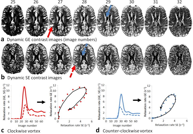
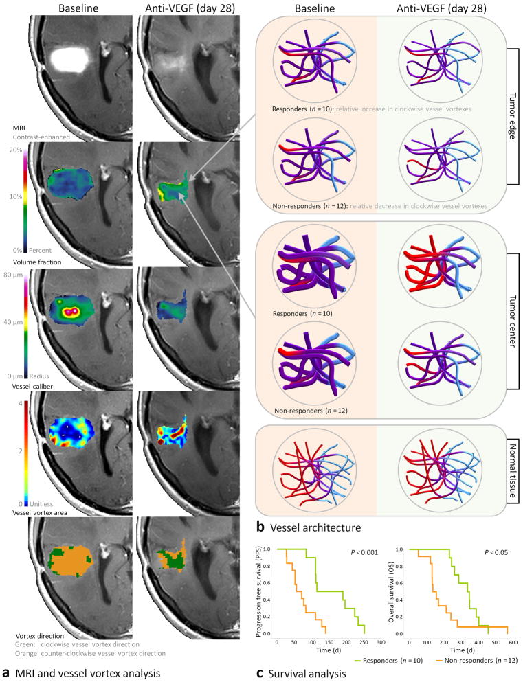
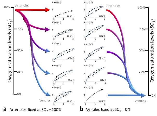
Measurement of vessel caliber by magnetic resonance imaging (MRI) is a valuable technique for in vivo monitoring of hemodynamic status and vascular development, especially in the brain. Here, we introduce a new paradigm in MRI termed vessel architectural imaging (VAI) that exploits an overlooked temporal shift in the magnetic resonance signal, forming the basis for vessel caliber estimation, and show how this phenomenon can reveal new information on vessel type and function not assessed by any other noninvasive imaging technique. We also show how this biomarker can provide new biological insights into the treatment of patients with cancer. As an example, we demonstrate using VAI that anti-angiogenic therapy can improve microcirculation and oxygen saturation and reduce vessel calibers in patients with recurrent glioblastomas and, more crucially, that patients with these responses have prolonged survival. Thus, VAI has the potential to identify patients who would benefit from therapies.

摘要

磁共振成像(MRI)测量血管口径是一种用于活体监测血液动力学状态和血管发育的有价值的技术,特别是在大脑中。在这里,我们引入了一种新的 MRI 方法,称为血管结构成像(VAI),它利用磁共振信号中被忽视的时间偏移,为血管口径估计奠定了基础,并展示了这一现象如何揭示其他任何非侵入性成像技术都无法评估的血管类型和功能的新信息。我们还展示了这种生物标志物如何为癌症患者的治疗提供新的生物学见解。例如,我们通过 VAI 证明,抗血管生成治疗可以改善复发性胶质母细胞瘤患者的微循环和氧饱和度,并减少血管口径,更重要的是,具有这些反应的患者的存活时间延长。因此,VAI 有可能识别出受益于治疗的患者。

https://cdn.ncbi.nlm.nih.gov/pmc/blobs/3284/3769525/51a0d1f526bb/nihms436751f5.jpg
https://cdn.ncbi.nlm.nih.gov/pmc/blobs/3284/3769525/98132f030842/nihms436751f1.jpg
https://cdn.ncbi.nlm.nih.gov/pmc/blobs/3284/3769525/6adb4468303a/nihms436751f2.jpg
https://cdn.ncbi.nlm.nih.gov/pmc/blobs/3284/3769525/8eb92cd4b0e9/nihms436751f3.jpg
https://cdn.ncbi.nlm.nih.gov/pmc/blobs/3284/3769525/fc095acc0913/nihms436751f4.jpg
https://cdn.ncbi.nlm.nih.gov/pmc/blobs/3284/3769525/51a0d1f526bb/nihms436751f5.jpg

相似文献

1
Vessel architectural imaging identifies cancer patient responders to anti-angiogenic therapy.血管构筑成像可识别出接受抗血管生成治疗的癌症患者的应答者。
Nat Med. 2013 Sep;19(9):1178-83. doi: 10.1038/nm.3289. Epub 2013 Aug 18.
2
Cediranib: profile of a novel anti-angiogenic agent in patients with glioblastoma.西地尼布:胶质母细胞瘤患者新型抗血管生成药物的概况
Expert Opin Investig Drugs. 2009 Oct;18(10):1549-57. doi: 10.1517/13543780903183528.
3
Imaging biomarkers guided anti-angiogenic therapy for malignant gliomas.影像生物标志物指导的恶性脑胶质瘤抗血管生成治疗。
Neuroimage Clin. 2018 Jul 5;20:51-60. doi: 10.1016/j.nicl.2018.07.001. eCollection 2018.
4
Edema control by cediranib, a vascular endothelial growth factor receptor-targeted kinase inhibitor, prolongs survival despite persistent brain tumor growth in mice.西地尼布(一种靶向血管内皮生长因子受体的激酶抑制剂)对水肿的控制,尽管小鼠脑肿瘤持续生长,但仍能延长生存期。
J Clin Oncol. 2009 May 20;27(15):2542-52. doi: 10.1200/JCO.2008.19.9356. Epub 2009 Mar 30.
5
Increased survival of glioblastoma patients who respond to antiangiogenic therapy with elevated blood perfusion.抗血管生成治疗后血供增加的胶质母细胞瘤患者的生存率提高。
Cancer Res. 2012 Jan 15;72(2):402-7. doi: 10.1158/0008-5472.CAN-11-2464. Epub 2011 Nov 29.
6
Phase II study of cediranib, an oral pan-vascular endothelial growth factor receptor tyrosine kinase inhibitor, in patients with recurrent glioblastoma.西地尼布(cediranib)是一种口服的泛血管内皮生长因子受体酪氨酸激酶抑制剂,在复发性胶质母细胞瘤患者中的 II 期研究。
J Clin Oncol. 2010 Jun 10;28(17):2817-23. doi: 10.1200/JCO.2009.26.3988. Epub 2010 May 10.
7
Glioblastoma recurrence after cediranib therapy in patients: lack of "rebound" revascularization as mode of escape.西地尼布治疗后胶质母细胞瘤复发的患者:无“反弹”血管生成作为逃逸模式。
Cancer Res. 2011 Jan 1;71(1):19-28. doi: 10.1158/0008-5472.CAN-10-2602.
8
A "vascular normalization index" as potential mechanistic biomarker to predict survival after a single dose of cediranib in recurrent glioblastoma patients.“血管正常化指数”作为预测复发性胶质母细胞瘤患者单次服用西地尼布后生存率的潜在机制生物标志物。
Cancer Res. 2009 Jul 1;69(13):5296-300. doi: 10.1158/0008-5472.CAN-09-0814. Epub 2009 Jun 23.
9
Anti-Angiogenics: Their Role in the Treatment of Glioblastoma.抗血管生成药物:在胶质母细胞瘤治疗中的作用。
Oncol Res Treat. 2018;41(4):181-186. doi: 10.1159/000488258. Epub 2018 Mar 23.
10
Preoperative biomarkers of tumour vascularity are elevated in patients with glioblastoma multiforme.多形性胶质母细胞瘤患者术前肿瘤血管生成的生物标志物升高。
J Clin Neurosci. 2015 Nov;22(11):1802-8. doi: 10.1016/j.jocn.2015.06.013. Epub 2015 Aug 22.

引用本文的文献

1
Vascular architecture mapping reveals sex-specific changes in cerebral microvasculature with aging.血管结构图谱揭示了随着衰老大脑微血管中特定性别的变化。
Imaging Neurosci (Camb). 2024 Jan 11;2. doi: 10.1162/imag_a_00066. eCollection 2024.
2
Vessel architecture imaging reveals microvascular rarefaction and capillary-to-arteriole shift in cerebral small vessel disease.血管结构成像显示脑小血管病中存在微血管稀疏和毛细血管向小动脉的转变。
J Cereb Blood Flow Metab. 2025 Jul 31:271678X251358968. doi: 10.1177/0271678X251358968.
3
Temporal Shift When Comparing Contrast-Agent Concentration Curves Estimated Using Quantitative Susceptibility Mapping (QSM) and ΔR2*: The Association Between Vortex Parameters and Oxygen Extraction Fraction.

本文引用的文献

1
Vascular normalizing doses of antiangiogenic treatment reprogram the immunosuppressive tumor microenvironment and enhance immunotherapy.抗血管生成治疗的血管正常化剂量可重新编程免疫抑制性肿瘤微环境并增强免疫治疗。
Proc Natl Acad Sci U S A. 2012 Oct 23;109(43):17561-6. doi: 10.1073/pnas.1215397109. Epub 2012 Oct 8.
2
Causes, consequences, and remedies for growth-induced solid stress in murine and human tumors.生长诱导的固体应力在鼠类和人类肿瘤中的原因、后果和补救措施。
Proc Natl Acad Sci U S A. 2012 Sep 18;109(38):15101-8. doi: 10.1073/pnas.1213353109. Epub 2012 Aug 29.
3
Increased survival of glioblastoma patients who respond to antiangiogenic therapy with elevated blood perfusion.
使用定量磁化率成像(QSM)和ΔR2*估计对比剂浓度曲线时的时间偏移:涡流参数与氧摄取分数之间的关联。
Tomography. 2025 Apr 9;11(4):46. doi: 10.3390/tomography11040046.
4
Mechanical forces in the tumor microenvironment: roles, pathways, and therapeutic approaches.肿瘤微环境中的机械力:作用、途径及治疗方法。
J Transl Med. 2025 Mar 12;23(1):313. doi: 10.1186/s12967-025-06306-8.
5
The Road to a Realistic 3D Model for Estimating R and R* Relaxation Versus Gd-DTPA Concentration in Whole Blood and Brain Tumor Vasculature.构建用于估计全血和脑肿瘤血管中R及R*弛豫与钆喷酸葡胺浓度关系的逼真三维模型之路
NMR Biomed. 2025 Jan;38(1):e5308. doi: 10.1002/nbm.5308.
6
Single-shot echo planar time-resolved imaging for multi-echo functional MRI and distortion-free diffusion imaging.用于多回波功能磁共振成像和无畸变扩散成像的单次激发回波平面时间分辨成像。
Magn Reson Med. 2025 Mar;93(3):993-1013. doi: 10.1002/mrm.30327. Epub 2024 Oct 20.
7
"Synthetic" DSC Perfusion MRI with Adjustable Acquisition Parameters in Brain Tumors Using Dynamic Spin-and-Gradient-Echo Echoplanar Imaging.使用动态自旋和梯度回波平面回波成像技术对脑肿瘤进行具有可调采集参数的“合成”DSC灌注MRI
AJNR Am J Neuroradiol. 2025 Feb 3;46(2):311-320. doi: 10.3174/ajnr.A8475.
8
Targeting the tumour vasculature: from vessel destruction to promotion.靶向肿瘤血管:从血管破坏到促进。
Nat Rev Cancer. 2024 Oct;24(10):655-675. doi: 10.1038/s41568-024-00736-0. Epub 2024 Aug 29.
9
Whole-Brain Vascular Architecture Mapping Identifies Region-Specific Microvascular Profiles In Vivo.全脑血管结构图谱描绘技术可在体识别特定区域的微血管特征。
AJNR Am J Neuroradiol. 2024 Sep 9;45(9):1346-1354. doi: 10.3174/ajnr.A8344.
10
Modulating cancer mechanopathology to restore vascular function and enhance immunotherapy.调节癌症机制病理学以恢复血管功能并增强免疫疗法。
Cell Rep Med. 2024 Jul 16;5(7):101626. doi: 10.1016/j.xcrm.2024.101626. Epub 2024 Jun 28.
抗血管生成治疗后血供增加的胶质母细胞瘤患者的生存率提高。
Cancer Res. 2012 Jan 15;72(2):402-7. doi: 10.1158/0008-5472.CAN-11-2464. Epub 2011 Nov 29.
4
Normalization of the vasculature for treatment of cancer and other diseases.血管正常化治疗癌症和其他疾病。
Physiol Rev. 2011 Jul;91(3):1071-121. doi: 10.1152/physrev.00038.2010.
5
Targeting hypoxia in cancer therapy.针对癌症治疗中的缺氧。
Nat Rev Cancer. 2011 Jun;11(6):393-410. doi: 10.1038/nrc3064.
6
Molecular mechanisms and clinical applications of angiogenesis.血管生成的分子机制与临床应用。
Nature. 2011 May 19;473(7347):298-307. doi: 10.1038/nature10144.
7
The shunt problem: control of functional shunting in normal and tumour vasculature.分流问题:正常和肿瘤血管中功能性分流的控制。
Nat Rev Cancer. 2010 Aug;10(8):587-93. doi: 10.1038/nrc2895. Epub 2010 Jul 15.
8
Phase II study of cediranib, an oral pan-vascular endothelial growth factor receptor tyrosine kinase inhibitor, in patients with recurrent glioblastoma.西地尼布(cediranib)是一种口服的泛血管内皮生长因子受体酪氨酸激酶抑制剂,在复发性胶质母细胞瘤患者中的 II 期研究。
J Clin Oncol. 2010 Jun 10;28(17):2817-23. doi: 10.1200/JCO.2009.26.3988. Epub 2010 May 10.
9
A fully automated method for quantitative cerebral hemodynamic analysis using DSC-MRI.一种使用 DSC-MRI 进行定量脑血流动力学分析的全自动方法。
J Cereb Blood Flow Metab. 2010 May;30(5):1066-78. doi: 10.1038/jcbfm.2010.4. Epub 2010 Jan 20.
10
Characterization of tumor angiogenesis in rat brain using iron-based vessel size index MRI in combination with gadolinium-based dynamic contrast-enhanced MRI.使用基于铁的血管大小指数磁共振成像结合基于钆的动态对比增强磁共振成像对大鼠脑肿瘤血管生成进行表征。
J Cereb Blood Flow Metab. 2009 Oct;29(10):1714-26. doi: 10.1038/jcbfm.2009.86. Epub 2009 Jul 8.