Department of Genetics and Developmental Biology, College of Life Science and Technology,Huazhong University of Science and Technology, Wuhan, Hubei, China.
PLoS One. 2013 Aug 19;8(8):e72015. doi: 10.1371/journal.pone.0072015. eCollection 2013.
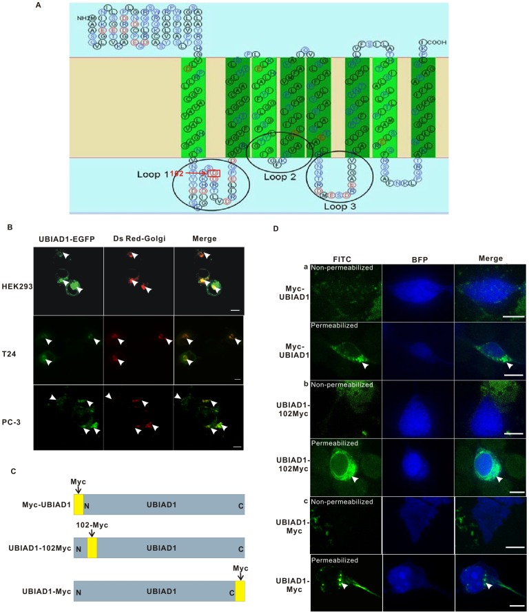
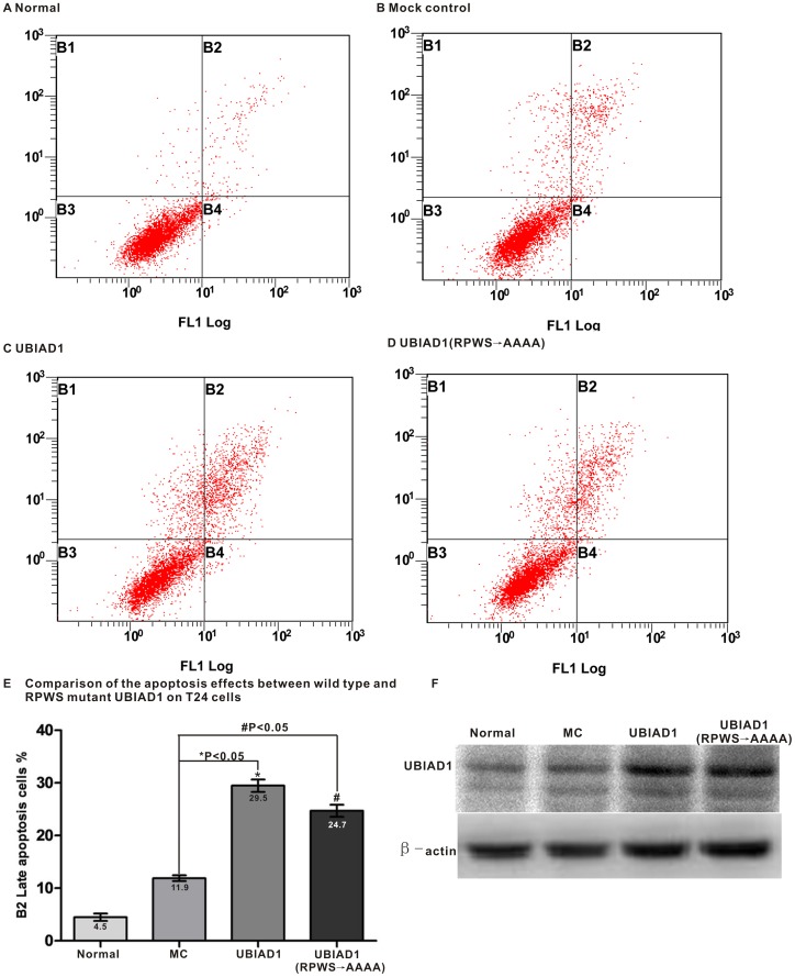
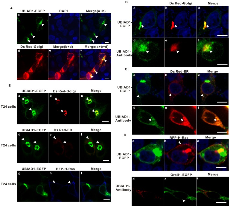
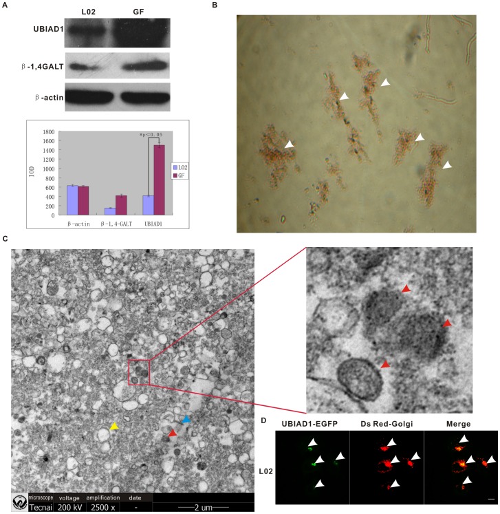
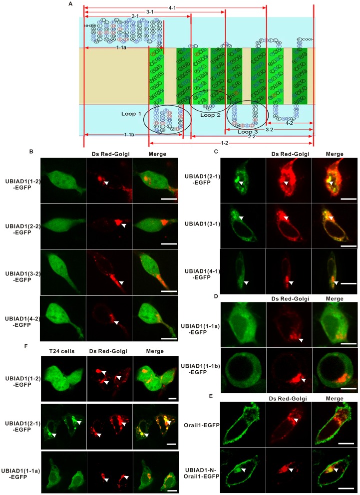
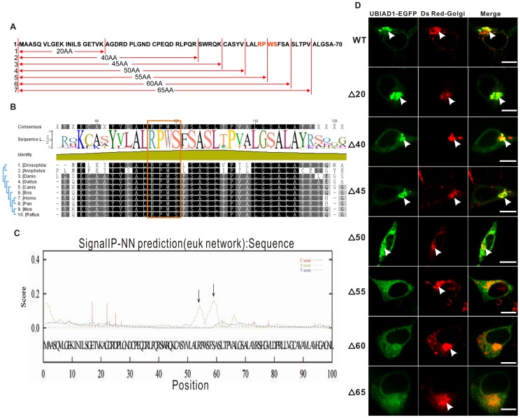
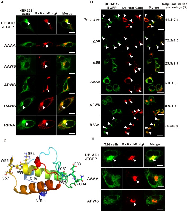
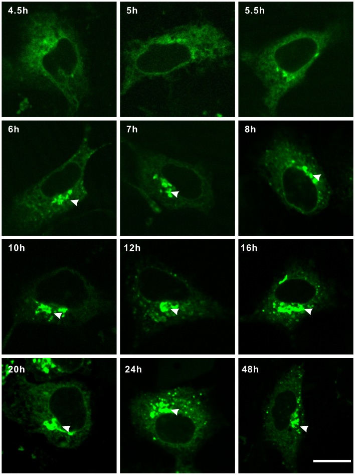
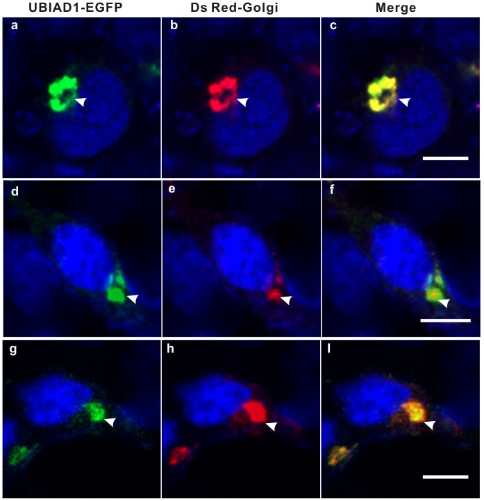
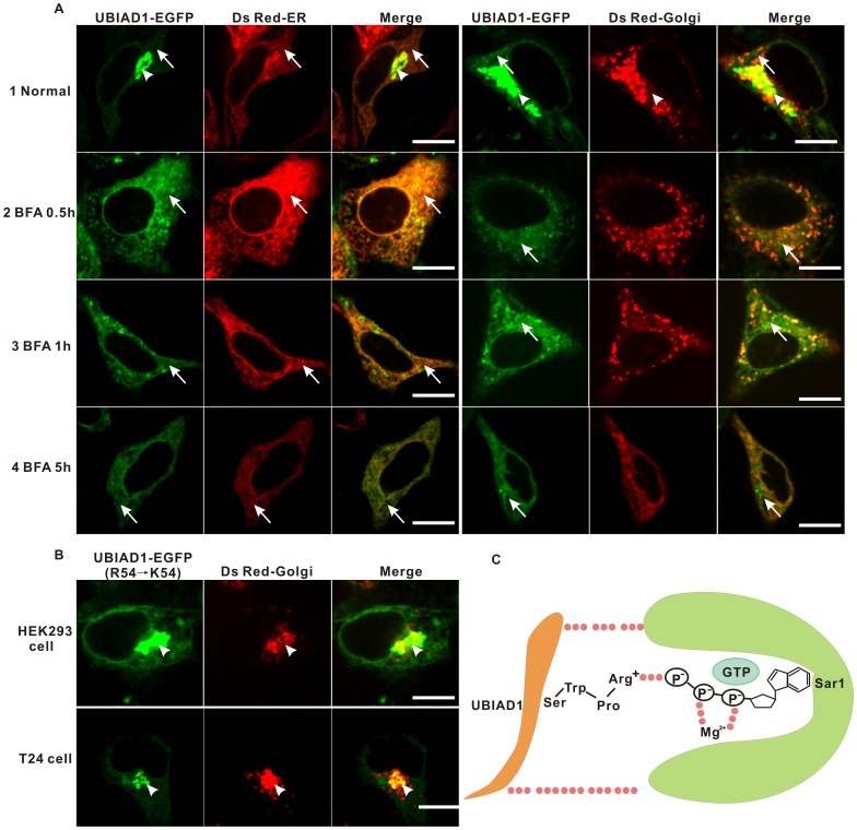
UBIAD1 plays critical roles in physiology including vitamin K and CoQ10 biosynthesis as well as pathophysiology including dyslipimedia-induced SCD (Schnyder's corneal dystrophy), Parkinson's disease, cardiovascular disease and bladder carcinoma. Since the subcellular localization of UBIAD1 varies in different cell types, characterization of the exact subcellular localization of UBIAD1 in specific human disease is vital for understanding its molecular mechanism. As UBIAD1 suppresses bladder carcinoma, we studied its subcellular localization in human bladder carcinoma cell line T24. Since fluorescent images of UBIAD1-EGFP in T24, human prostate cancer cell line PC-3, human embryonic kidney cell line HEK293 and human hepatocyte cell line L02 are similar, these four cell lines were used for present study. Using a combination of fluorescent microscopy and immunohistochemistry, it was found that UBIAD1 localized on the Golgi and endoplasmic reticulum (ER), but not on the plasma membrane, of T24 and HEK293 cells. Using scanning electron microscopy and western blot analysis, we found that UBIAD1 is enriched in the Golgi fraction extracted from the L02 cells, verifying the Golgi localization of UBAID1. Site-directed mutagenesis showed that the RPWS motif, which forms an Arginine finger on the UBIAD1 N terminus, serves as the Golgi retention signal. With both cycloheximide and brefeldin A inhibition assays, it was shown that UBIAD1 may be transported from the endoplasmic reticulum (ER) to the Golgi by a COPII-mediated mechanism. Based upon flow cytometry analysis, it is shown that mutation of the RPWS motif reduced the UBIAD1-induced apoptosis of T24 cells, indicating that the proper Golgi localization of UBIAD1 influences its tumor suppressant activity. This study paves the way for further understanding the molecular mechanism of UBIAD1 in human diseases.
UBIAD1 在生理学中发挥着关键作用,包括维生素 K 和 CoQ10 的生物合成,以及病理生理学,包括脂代谢异常诱导的 SCD(施奈德角膜营养不良)、帕金森病、心血管疾病和膀胱癌。由于 UBIAD1 的亚细胞定位在不同的细胞类型中有所不同,因此,确定 UBIAD1 在特定人类疾病中的精确亚细胞定位对于理解其分子机制至关重要。由于 UBIAD1 抑制膀胱癌,我们研究了其在人膀胱癌 T24 细胞系中的亚细胞定位。由于 UBIAD1-EGFP 在 T24、人前列腺癌细胞系 PC-3、人胚肾细胞系 HEK293 和人肝细胞系 L02 中的荧光图像相似,因此本研究使用了这四种细胞系。通过荧光显微镜和免疫组织化学结合的方法,发现 UBIAD1 定位于 T24 和 HEK293 细胞的高尔基体和内质网(ER),而不在质膜上。通过扫描电子显微镜和 Western blot 分析,我们发现 UBIAD1 富集在从 L02 细胞提取的高尔基体部分中,验证了 UBAID1 的高尔基体定位。定点突变显示,形成 UBIAD1 N 端精氨酸指的 RPWS 基序是高尔基体保留信号。通过环己酰亚胺和布雷菲德菌素 A 抑制实验表明,UBIAD1 可能通过 COPII 介导的机制从内质网(ER)转运到高尔基体。基于流式细胞术分析,表明 RPWS 基序的突变降低了 UBIAD1 诱导的 T24 细胞凋亡,表明 UBIAD1 的正确高尔基体定位影响其肿瘤抑制活性。这项研究为进一步了解 UBIAD1 在人类疾病中的分子机制铺平了道路。