Department of Tropical Nutrition and Food Science, Faculty of Tropical Medicine, Mahidol University Bangkok, Thailand.
PLoS One. 2013 Aug 21;8(8):e70386. doi: 10.1371/journal.pone.0070386. eCollection 2013.
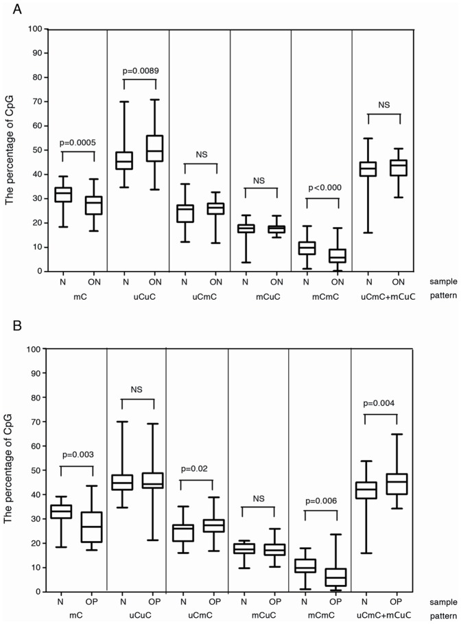
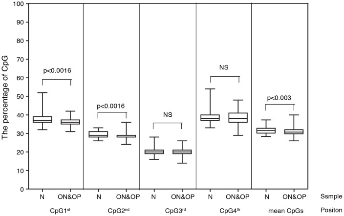
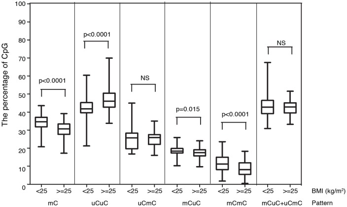
A decrease in genomic methylation commonly occurs in aging cells; however, whether this epigenetic modification leads to age-related phenotypes has not been evaluated. Alu elements are the major interspersed repetitive DNA elements in humans that lose DNA methylation in aging individuals. Alu demethylation in blood cells starts at approximately 40 years of age, and the degree of Alu hypomethylation increases with age. Bone mass is lost with aging, particularly in menopausal women with lower body mass. Consequently, osteoporosis is commonly found in thin postmenopausal women. Here, we correlated the Alu methylation level of blood cells with bone density in 323 postmenopausal women. Alu hypomethylation was associated with advanced age and lower bone mass density, (P<0.05). The association between the Alu methylation level and bone mass was independent of age, body mass, and body fat, with an odds ratio [1] = 0.4316 (0.2087-0.8927). Individuals of the same age with osteopenia, osteoporosis, and a high body mass index have lower Alu methylation levels (P = 0.0005, 0.003, and ≤0.0001, respectively). Finally, when comparing individuals with the same age and body mass, Alu hypomethylation was observed in individuals with lower bone mass (P<0.0001). In conclusion, there are positive correlations between Alu hypomethylation in blood cells and several age-related phenotypes in bone and body fat. Therefore, reduced global methylation may play a role in the systemic senescence process. Further evaluation of Alu hypomethylation may clarify the epigenetic regulation of osteoporosis in post-menopausal women.
基因组甲基化水平普遍降低发生在衰老细胞中;然而,这种表观遗传修饰是否导致与年龄相关的表型尚未得到评估。Alu 元件是人类中主要的散在重复 DNA 元件,在衰老个体中失去 DNA 甲基化。血细胞中的 Alu 去甲基化始于大约 40 岁,Alu 低甲基化程度随年龄增加而增加。随着年龄的增长,骨量会丢失,尤其是在体重较低的绝经后妇女中。因此,骨质疏松症常见于瘦绝经后妇女。在这里,我们将 323 名绝经后妇女的血细胞 Alu 甲基化水平与骨密度相关联。Alu 低甲基化与年龄较大和较低的骨密度密度相关(P<0.05)。Alu 甲基化水平与骨量之间的关联独立于年龄、体重和体脂肪,优势比 [1]为 0.4316(0.2087-0.8927)。患有骨质疏松症、骨质疏松症和高体重指数的同年龄个体的 Alu 甲基化水平较低(P=0.0005、0.003 和≤0.0001)。最后,当比较同年龄和体重的个体时,在骨量较低的个体中观察到 Alu 低甲基化(P<0.0001)。总之,血细胞中的 Alu 低甲基化与骨骼和体脂肪中的几种与年龄相关的表型之间存在正相关。因此,整体甲基化减少可能在全身衰老过程中发挥作用。进一步评估 Alu 低甲基化可能会阐明绝经后妇女骨质疏松症的表观遗传调控。