Department of Oncology, San Raffaele Scientific Institute, Milan, Italy.
PLoS One. 2013 Aug 26;8(8):e71613. doi: 10.1371/journal.pone.0071613. eCollection 2013.
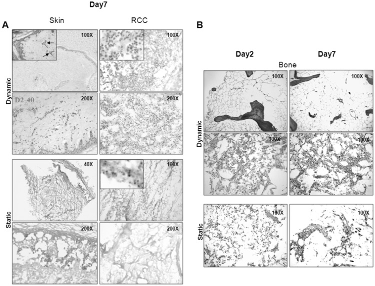
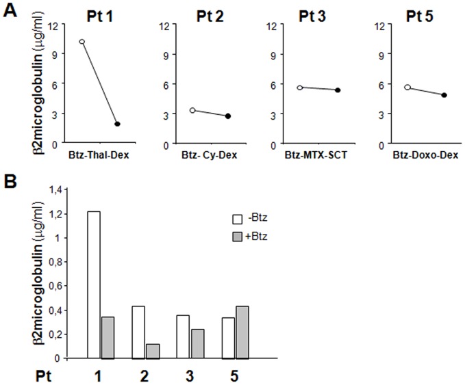
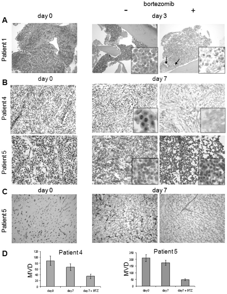
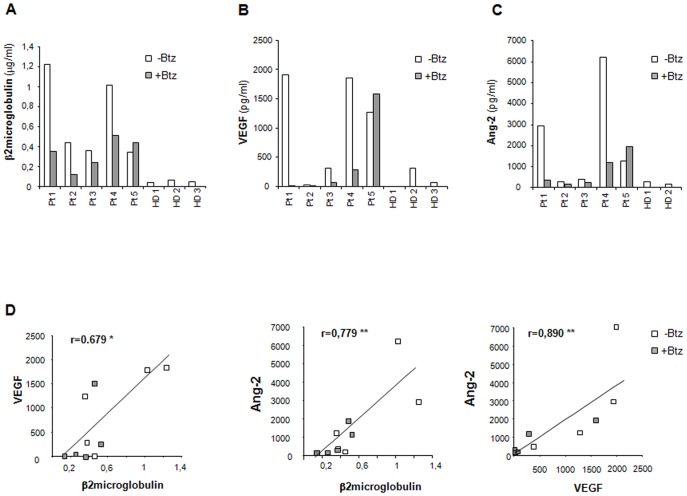
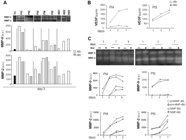
Three-dimensional (3-D) culture models are emerging as invaluable tools in tumor biology, since they reproduce tissue-specific structural features and cell-cell interactions more accurately than conventional 2-D cultures. Multiple Myeloma, which depends on myeloma cell-Bone Marrow microenvironment interactions for development and response to drugs, may particularly benefit from such an approach. An innovative 3-D dynamic culture model based on the use of the RCCS™ Bioreactor was developed to allow long-term culture of myeloma tissue explants. This model was first validated with normal and pathological explants, then applied to tissues from myeloma patients. In all cases, histological examination demonstrated maintenance of viable myeloma cells inside their native microenvironment, with an overall well preserved histo-architecture including bone lamellae and vessels. This system was then successfully applied to evaluate the cytotoxic effects exerted by the proteasome inhibitor Bortezomib not only on myeloma cells but also on angiogenic vessels. Moreover, as surrogate markers of specialized functions expressed by myeloma cells and microenvironment, β2 microglobulin, VEGF and Angiopoietin-2 levels, as well as Matrix Metalloproteases activity, were evaluated in supernatants from 3D cultures and their levels reflected the effects of Bortezomib treatment. Notably, determination of β2 microglobulin levels in supernatants from Bortezomib-treated samples and in patients'sera following Bortezomib-based therapies disclosed an overall concordance in the response to the drug ex vivo and in vivo. Our findings indicate, as a proof of principle, that 3-D, RCCS™ bioreactor-based culture of tissue explants can be exploited for studying myeloma biology and for a pre-clinical approach to patient-targeted therapy.
三维(3-D)培养模型作为肿瘤生物学中极具价值的工具正在兴起,因为它们比传统的 2-D 培养更准确地复制组织特异性结构特征和细胞间相互作用。多发性骨髓瘤依赖于骨髓瘤细胞-骨髓微环境相互作用的发展和对药物的反应,可能特别受益于这种方法。开发了一种基于 RCCS™生物反应器使用的创新的 3-D 动态培养模型,以允许骨髓瘤组织外植体的长期培养。该模型首先用正常和病理外植体进行验证,然后应用于骨髓瘤患者的组织。在所有情况下,组织学检查都证明了活骨髓瘤细胞在其天然微环境中的维持,具有总体保存良好的组织架构,包括骨板层和血管。然后,该系统成功地应用于评估蛋白酶体抑制剂硼替佐米不仅对骨髓瘤细胞而且对血管生成血管的细胞毒性作用。此外,作为骨髓瘤细胞和微环境表达的特殊功能的替代标志物,β2 微球蛋白、VEGF 和血管生成素-2 水平以及基质金属蛋白酶活性,在 3D 培养物的上清液中进行了评估,并且它们的水平反映了硼替佐米处理的效果。值得注意的是,在硼替佐米处理的样本的上清液中和硼替佐米治疗后的患者血清中测定β2 微球蛋白水平,显示了药物在体外和体内反应的总体一致性。我们的研究结果表明,作为原理证明,组织外植体的 3-D、RCCS™生物反应器培养可用于研究骨髓瘤生物学和进行针对患者的治疗的临床前方法。