Department of Physiology, University of Arizona, Tucson, Arizona, United States of America.
PLoS One. 2013 Aug 21;8(8):e72080. doi: 10.1371/journal.pone.0072080. eCollection 2013.
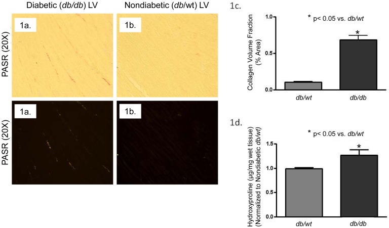
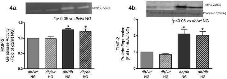
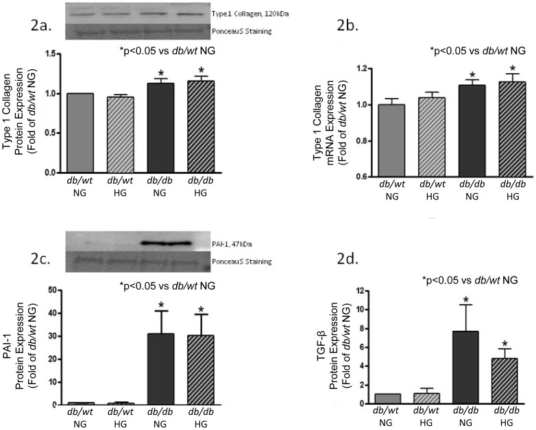
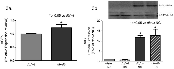
Cardiovascular complications are a leading cause of death in patients with type 2 diabetes mellitus (T2DM). Diastolic dysfunction is one of the earliest manifestations of diabetes-induced changes in left ventricular (LV) function, and results from a reduced rate of relaxation and increased stiffness. The mechanisms responsible for increased stiffness are not completely understood. Chronic hyperglycemia, advanced glycation endproducts (AGEs), and increased levels of proinflammatory and profibrotic cytokines are molecular pathways known to be involved in regulating extracellular matrix (ECM) synthesis and accumulation resulting in increased LV diastolic stiffness. Experiments were conducted using a genetically-induced mouse model of T2DM generated by a point mutation in the leptin receptor resulting in nonfunctional leptin receptors (db/db murine model). This study correlated changes in LV ECM and stiffness with alterations in basal activation of signaling cascades and expression of profibrotic markers within primary cultures of cardiac fibroblasts from diabetic (db/db) mice with nondiabetic (db/wt) littermates as controls. Primary cultures of cardiac fibrobroblasts were maintained in 25 mM glucose (hyperglycemic-HG; diabetic db/db) media or 5 mM glucose (normoglycemic-NG, nondiabetic db/wt) media. The cells then underwent a 24-hour exposure to their opposite (NG; diabetic db/db) media or 5 mM glucose (HG, nondiabetic db/wt) media. Protein analysis demonstrated significantly increased expression of type I collagen, TIMP-2, TGF-β, PAI-1 and RAGE in diabetic db/db cells as compared to nondiabetic db/wt, independent of glucose media concentration. This pattern of protein expression was associated with increased LV collagen accumulation, myocardial stiffness and LV diastolic dysfunction. Isolated diabetic db/db fibroblasts were phenotypically distinct from nondiabetic db/wt fibroblasts and exhibited a profibrotic phenotype in normoglycemic conditions.
心血管并发症是 2 型糖尿病(T2DM)患者死亡的主要原因。舒张功能障碍是糖尿病引起的左心室(LV)功能变化的最早表现之一,其原因是舒张期松弛率降低和僵硬度增加。导致僵硬度增加的机制尚未完全阐明。慢性高血糖、晚期糖基化终产物(AGEs)和促炎及促纤维化细胞因子水平升高,这些分子途径已知参与调节细胞外基质(ECM)的合成和积累,导致 LV 舒张僵硬增加。实验使用一种通过瘦素受体点突变产生的 2 型糖尿病遗传诱导小鼠模型进行,该突变导致瘦素受体无功能(db/db 鼠模型)。本研究将 LV ECM 和僵硬度的变化与糖尿病(db/db)小鼠与非糖尿病(db/wt)同窝仔鼠心脏成纤维细胞原代培养中信号转导通路基础激活和促纤维化标志物表达的改变相关联。心脏成纤维细胞原代培养物在 25 mM 葡萄糖(高血糖-HG;糖尿病 db/db)培养基或 5 mM 葡萄糖(正常血糖-NG,非糖尿病 db/wt)培养基中维持。然后,细胞接受 24 小时相反(NG;糖尿病 db/db)培养基或 5 mM 葡萄糖(HG,非糖尿病 db/wt)培养基的处理。蛋白分析表明,与非糖尿病 db/wt 相比,糖尿病 db/db 细胞中 I 型胶原、TIMP-2、TGF-β、PAI-1 和 RAGE 的表达显著增加,而与葡萄糖培养基浓度无关。这种蛋白表达模式与 LV 胶原积累增加、心肌僵硬度增加和 LV 舒张功能障碍有关。与非糖尿病 db/wt 成纤维细胞相比,分离的糖尿病 db/db 成纤维细胞具有明显不同的表型,并在正常血糖条件下表现出促纤维化表型。