Centre for Comparative and Clinical Anatomy, University of Bristol, Southwell Street, Bristol BS2 8EJ, UK.
Int J Mol Sci. 2013 Aug 29;14(9):17729-43. doi: 10.3390/ijms140917729.
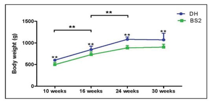
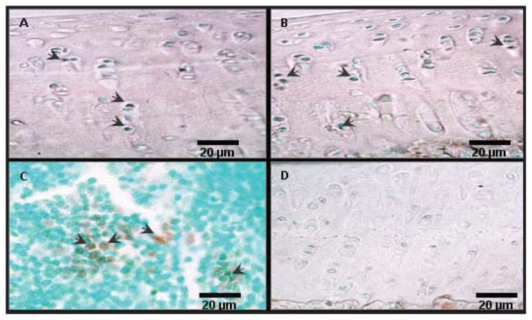
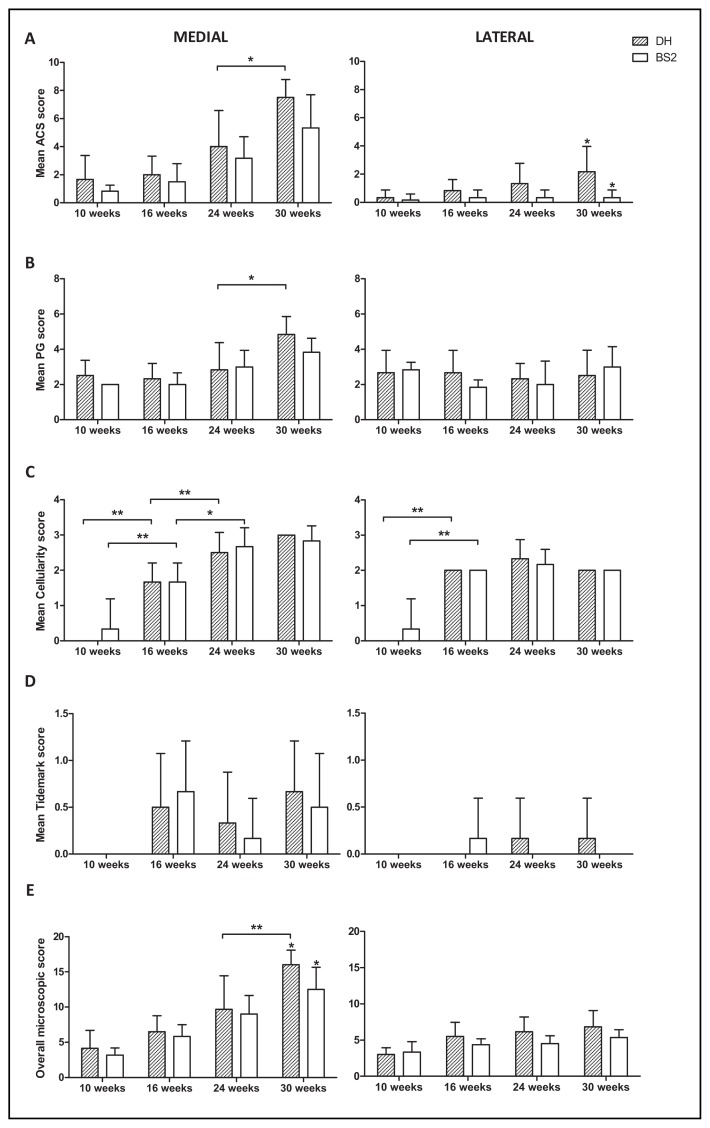
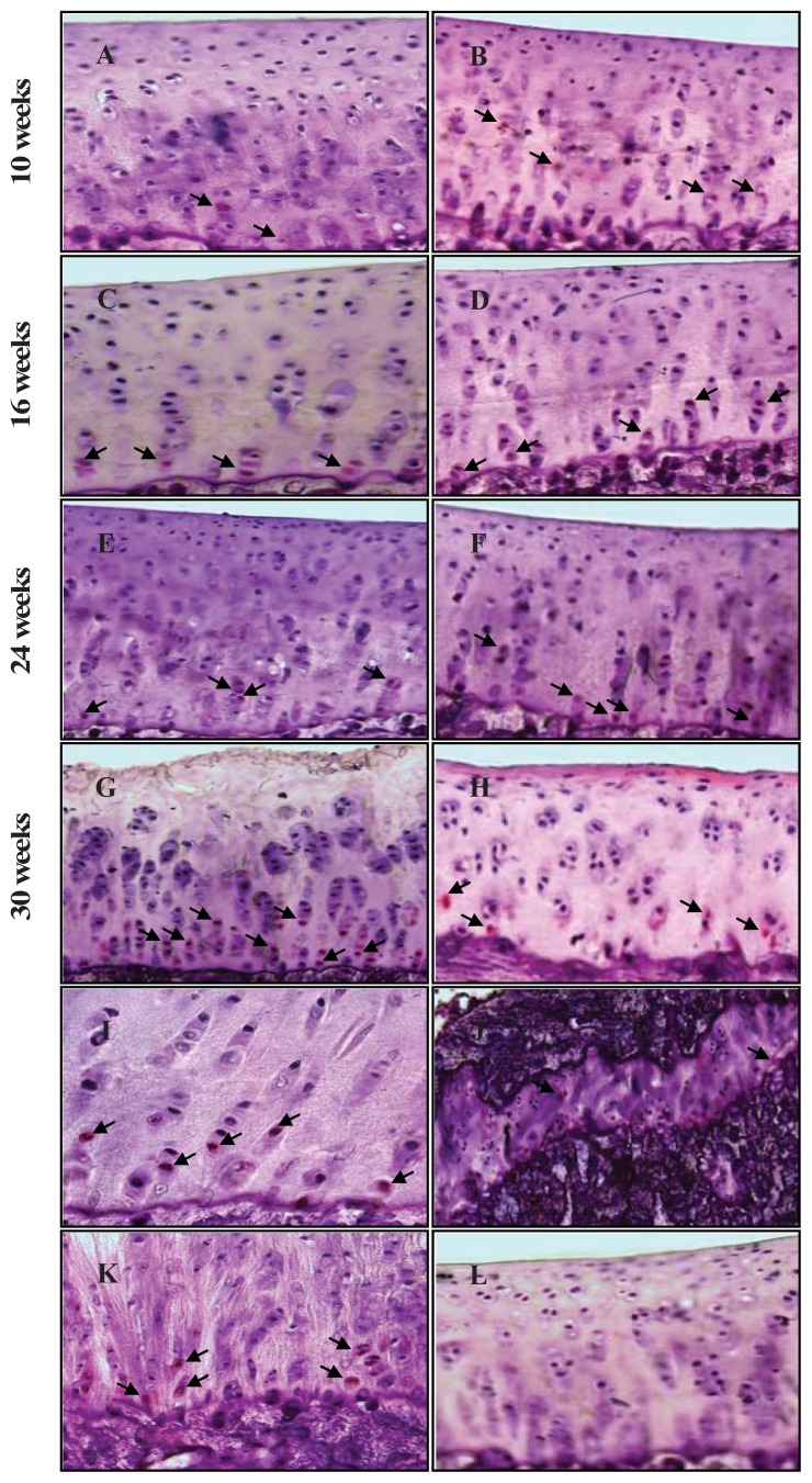
Osteoarthritis (OA) is the most common joint disease characterised by degradation of articular cartilage and bone remodelling. For almost a decade chondrocyte apoptosis has been investigated as a possible mechanism of cartilage damage in OA, but its precise role in initiation and/or progression of OA remains to the determined. The aim of this study is to determine the role of chondrocyte apoptosis in spontaneous animal models of OA. Right tibias from six male Dunkin Hartley (DH) and Bristol Strain 2 (BS2) guinea pigs were collected at 10, 16, 24 and 30 weeks of age. Fresh-frozen sections of tibial epiphysis were microscopically scored for OA, and immunostained with caspase-3 and TUNEL for apoptotic chondrocytes. The DH strain had more pronounced cartilage damage than BS2, especially at 30 weeks. At this time point, the apoptotic chondrocytes were largely confined to the deep zone of articular cartilage (AC) with a greater percentage in the medial side of DH than BS2 (DH: 5.7%, 95% CI: 4.2-7.2), BS2: 4.8%, 95% CI: 3.8-5.8), p > 0.05). DH had a significant progression of chondrocyte death between 24 to 30 weeks during which time significant changes were observed in AC fibrillation, proteoglycan depletion and overall microscopic OA score. A strong correlation (p ≤ 0.01) was found between chondrocyte apoptosis and AC fibrillation (r = 0.3), cellularity (r = 0.4) and overall microscopic OA scores (r = 0.4). Overall, the rate of progression in OA and apoptosis over the study period was greater in the DH (versus BS2) and the medial AC (versus lateral). Chondrocyte apoptosis was higher at the later stage of OA development when the cartilage matrix was hypocellular and highly fibrillated, suggesting that chondrocyte apoptosis is a late event in OA.
骨关节炎(OA)是最常见的关节疾病,其特征为关节软骨降解和骨重塑。近十年来,软骨细胞凋亡已被研究为 OA 软骨损伤的可能机制,但它在 OA 的起始和/或进展中的确切作用仍有待确定。本研究旨在确定软骨细胞凋亡在 OA 自发动物模型中的作用。在 10、16、24 和 30 周龄时,收集 6 只雄性 Dunkin Hartley(DH)和 Bristol Strain 2(BS2)豚鼠的右胫骨。通过显微镜对胫骨骺端进行 OA 评分,并通过 caspase-3 和 TUNEL 免疫染色检测凋亡的软骨细胞。DH 品系比 BS2 品系有更明显的软骨损伤,尤其是在 30 周时。在这一时间点,凋亡的软骨细胞主要局限于关节软骨(AC)的深层区,DH 的内侧比 BS2 的百分比更高(DH:5.7%,95%CI:4.2-7.2),BS2:4.8%,95%CI:3.8-5.8),p>0.05)。DH 在 24 周至 30 周之间,软骨细胞死亡呈显著进展,在此期间,AC 纤维化、糖胺聚糖耗竭和整体微观 OA 评分均发生显著变化。软骨细胞凋亡与 AC 纤维化(r=0.3)、细胞密度(r=0.4)和整体微观 OA 评分(r=0.4)之间存在很强的相关性(p≤0.01)。总的来说,在 DH(与 BS2 相比)和内侧 AC(与外侧相比)中,OA 和凋亡在研究期间的进展速度更快。在软骨基质细胞稀少和高度纤维化的 OA 晚期,软骨细胞凋亡更高,这表明软骨细胞凋亡是 OA 的晚期事件。