Neuroimmunology Research, R&D-31, Portland Veterans Affairs Medical Center, 3710 SW US Veterans Hospital Road, Portland, OR 97239, USA.
J Neuroinflammation. 2013 Sep 9;10:111. doi: 10.1186/1742-2094-10-111.
Stroke severity is worsened by recruitment of inflammatory immune cells into the brain. This process depends in part on T cell activation, in which the B7 family of co-stimulatory molecules plays a pivotal role. Previous studies demonstrated more severe infarcts in mice lacking programmed death-1 (PD-1), a member of the B7 family, thus implicating PD-1 as a key factor in limiting stroke severity. The purpose of this study was to determine if this protective effect of PD-1 involves either of its ligands, PD-L1 or PD-L2.
Central nervous system (CNS) inflammation and infarct volume were evaluated in male PD-L1 and PD-L2 knockout (-/-) mice undergoing 60 minutes of middle cerebral artery occlusion (MCAO) followed by 96 hours of reperfusion and compared to wild-type (WT) C57BL/6J mice.
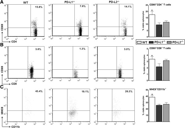
PD-L1-/- and PD-L2-/- mice had smaller total infarct volumes compared to WT mice. The PD-L1-/- and to a lesser extent PD-L2-/- mice had reduced levels of proinflammatory activated microglia and/or infiltrating monocytes and CD4+ T cells in the ischemic hemispheres. There was a reduction in ischemia-related splenic atrophy accompanied by lower activation status of splenic T cells and monocytes in the absence of PD-L1, suggesting a pathogenic rather than a regulatory role for both PD-1 ligands (PD-Ls). Suppressor T cells (IL-10-producing CD8+CD122+ T cells) trafficked to the brain in PD-L1-/- mice and there was decreased expression of CD80 on splenic antigen-presenting cells (APCs) as compared to the WT and PD-L2-/- mice.
Our novel observations are the first to implicate PD-L1 involvement in worsening outcome of experimental stroke. The presence of suppressor T cells in the right MCAO-inflicted hemisphere in mice lacking PD-L1 implicates these cells as possible key contributors for controlling adverse effects of ischemia. Increased expression of CD80 on APCs in WT and PD-L2-/- mice suggests an overriding interaction leading to T cell activation. Conversely, low CD80 expression by APCs, along with increased PD-1 and PD-L2 expression in PD-L1-/- mice suggests alternative T cell signaling pathways, leading to a suppressor phenotype. These results suggest that agents (for example antibodies) that can target and neutralize PD-L1/2 may have therapeutic potential for treatment of human stroke.
炎症免疫细胞被募集到大脑中会加重中风的严重程度。这个过程部分依赖于 T 细胞的激活,其中 B7 家族共刺激分子起着关键作用。先前的研究表明,程序性死亡受体 1(PD-1)缺失的小鼠(PD-1 是 B7 家族的一员)梗死更严重,因此 PD-1 被认为是限制中风严重程度的关键因素。本研究旨在确定 PD-1 的这种保护作用是否涉及它的配体 PD-L1 或 PD-L2。
对雄性 PD-L1 和 PD-L2 敲除(-/-)小鼠进行 60 分钟大脑中动脉闭塞(MCAO)后 96 小时再灌注,评估中枢神经系统(CNS)炎症和梗死体积,并与野生型(WT)C57BL/6J 小鼠进行比较。
与 WT 小鼠相比,PD-L1-/-和 PD-L2-/-小鼠的总梗死体积更小。PD-L1-/-和 PD-L2-/-小鼠的缺血半球中促炎激活的小胶质细胞和/或浸润的单核细胞和 CD4+T 细胞水平降低。在缺乏 PD-L1 的情况下,与缺血相关的脾脏萎缩减少,同时脾脏 T 细胞和单核细胞的激活状态降低,这表明 PD-1 配体(PD-Ls)具有致病性而不是调节作用。IL-10 产生的 CD8+CD122+T 细胞在 PD-L1-/-小鼠中迁移到大脑,与 WT 和 PD-L2-/-小鼠相比,脾抗原呈递细胞(APC)上 CD80 的表达降低。
我们的新发现首次表明 PD-L1 参与了实验性中风结局的恶化。在缺乏 PD-L1 的右 MCAO 损伤半球中存在抑制性 T 细胞表明这些细胞可能是控制缺血不良影响的关键因素。WT 和 PD-L2-/-小鼠中 APC 上 CD80 的高表达表明存在一种主导的相互作用,导致 T 细胞激活。相反,PD-L1-/-小鼠 APC 上 CD80 的低表达,加上 PD-1 和 PD-L2 的高表达,提示存在替代的 T 细胞信号通路,导致抑制表型。这些结果表明,针对 PD-L1/2 的药物(例如抗体)可能具有治疗人类中风的治疗潜力。