Huang Yimin, Hu Lin, Zhang Tingting, Zhong Hao, Zhou Jiajia, Liu Zhenbang, Wang Haibao, Guo Zhen, Chen Qianwang
Hefei National Laboratory for Physical Sciences at Microscale and Department of Materials Science & Engineering, University of Science and Technology of China, Hefei 230026, China.
Sci Rep. 2013;3:2647. doi: 10.1038/srep02647.
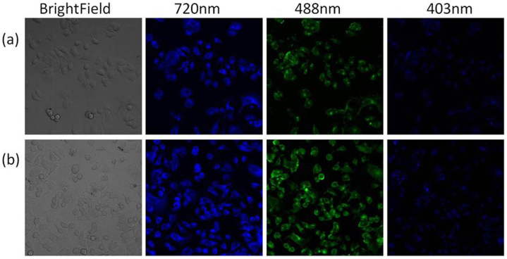
Nanoprobes with dual modal imaging of magnetic resonance imaging (MRI) and two-photon fluorescence (TPF) can serve as promising platforms for clinical diagnosis. A wide range of molecules and nanoparticles have been investigated as agents for contrast enhanced MRI and fluorescence imaging in cancer diagnosis. However, a single material with dual modal imaging of MRI and TPF is rarely reported. We found that Mn₃[Co(CN)₆]₂@SiO₂ nanocubes can serve as agents for both T₁- and T₂-weighted MRI, and TPF imaging. The nanocubes coated with silica to form Mn₃[Co(CN)₆]₂@SiO₂ core-shell nanocubes were readily internalized by cells without showing cytotoxicity. In vitro tests, the core-shell nanocubes display relatively high longitudinal (r₁) and transverse (r₂) relaxivities, they also manifest a remarkable T₁ and T₂ contrast effects at in-vivo imaging of internal organs in Mice. Moreover, the core-shell nanocubes could offer high-resolution cell fluorescence imaging by two-photon excitation (720 nm) or by conventional fluorescence with 403- or 488-nm excitation.
具有磁共振成像(MRI)和双光子荧光(TPF)双模态成像功能的纳米探针可作为临床诊断的有前景平台。已对多种分子和纳米颗粒作为癌症诊断中对比增强MRI和荧光成像的试剂进行了研究。然而,很少有报道单一材料同时具备MRI和TPF双模态成像功能。我们发现Mn₃[Co(CN)₆]₂@SiO₂纳米立方体可作为T₁加权和T₂加权MRI以及TPF成像的试剂。包覆二氧化硅形成Mn₃[Co(CN)₆]₂@SiO₂核壳纳米立方体的纳米立方体易于被细胞内化且无细胞毒性。体外测试中,核壳纳米立方体表现出相对较高的纵向(r₁)和横向(r₂)弛豫率,在小鼠体内器官成像中也表现出显著的T₁和T₂对比效果。此外,核壳纳米立方体可通过双光子激发(720 nm)或403-或488-nm激发的传统荧光提供高分辨率细胞荧光成像。