Oxford Centre for Clinical Magnetic Resonance Research, Division of Cardiovascular Medicine, Radcliffe Department of Medicine, University of Oxford, West Wing, Level 6, John Radcliffe Hospital, Oxford, UK.
Oxford Centre for Clinical Magnetic Resonance Research, Division of Cardiovascular Medicine, Radcliffe Department of Medicine, University of Oxford, West Wing, Level 6, John Radcliffe Hospital, Oxford, UK; Translational Gastroenterology Unit, University of Oxford, Level 5, John Radcliffe Hospital, Oxford, UK.
J Hepatol. 2014 Jan;60(1):69-77. doi: 10.1016/j.jhep.2013.09.002. Epub 2013 Sep 12.
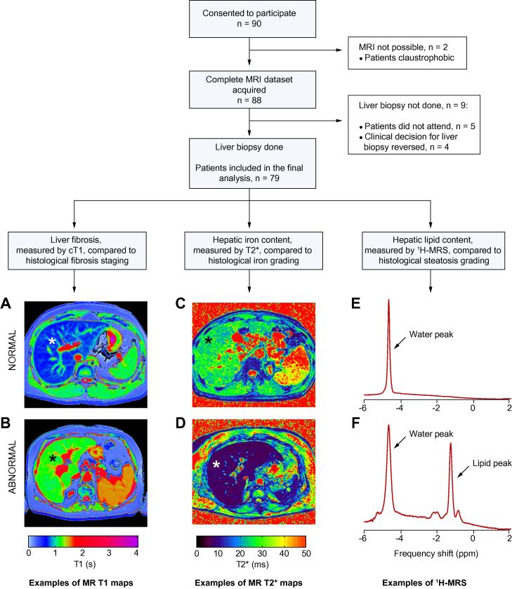
BACKGROUND & AIMS: With the increasing prevalence of liver disease worldwide, there is an urgent clinical need for reliable methods to diagnose and stage liver pathology. Liver biopsy, the current gold standard, is invasive and limited by sampling and observer dependent variability. In this study, we aimed to assess the diagnostic accuracy of a novel magnetic resonance protocol for liver tissue characterisation.
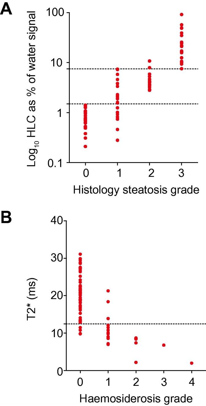
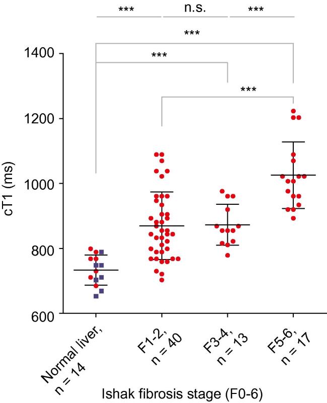
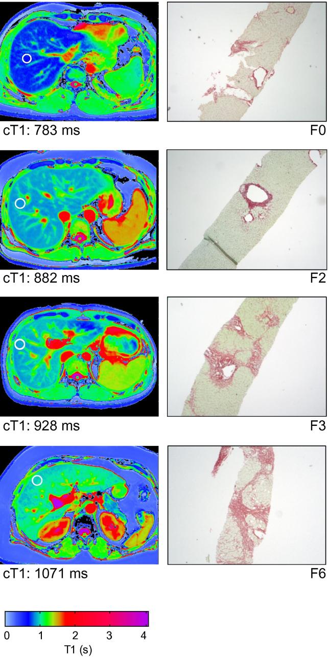
We conducted a prospective study comparing our magnetic resonance technique against liver biopsy. The individual components of the scanning protocol were T1 mapping, proton spectroscopy and T2* mapping, which quantified liver fibrosis, steatosis and haemosiderosis, respectively. Unselected adult patients referred for liver biopsy as part of their routine care were recruited. Scans performed prior to liver biopsy were analysed by physicians blinded to the histology results. The associations between magnetic resonance and histology variables were assessed. Receiver-operating characteristic analyses were also carried out.
Paired magnetic resonance and biopsy data were obtained in 79 patients. Magnetic resonance measures correlated strongly with histology (r(s)=0.68 p<0.0001 for fibrosis; r(s)=0.89 p<0.001 for steatosis; r(s)=-0.69 p<0.0001 for haemosiderosis). The area under the receiver operating characteristic curve was 0.94, 0.93, and 0.94 for the diagnosis of any degree of fibrosis, steatosis and haemosiderosis respectively.
The novel scanning method described here provides high diagnostic accuracy for the assessment of liver fibrosis, steatosis and haemosiderosis and could potentially replace liver biopsy for many indications. This is the first demonstration of a non-invasive test to differentiate early stages of fibrosis from normal liver.
随着全球肝病患病率的增加,临床迫切需要可靠的方法来诊断和分期肝脏病理。肝活检是目前的金标准,但具有侵袭性,且受到取样和观察者变异的限制。本研究旨在评估一种新的磁共振方案用于肝脏组织特征诊断的准确性。
我们进行了一项前瞻性研究,比较了我们的磁共振技术与肝活检。扫描方案的各个组成部分分别为 T1 映射、质子波谱和 T2*映射,分别定量纤维化、脂肪变性和血色素沉着症。我们招募了因常规护理而接受肝活检的未选择的成年患者。在对组织学结果不知情的情况下,由医师对扫描前的磁共振图像进行分析。评估磁共振和组织学变量之间的相关性,并进行了受试者工作特征分析。
79 例患者获得了配对的磁共振和活检数据。磁共振测量与组织学结果相关性较强(纤维化 r(s)=0.68,p<0.0001;脂肪变性 r(s)=0.89,p<0.001;血色素沉着症 r(s)=-0.69,p<0.0001)。受试者工作特征曲线下面积分别为 0.94、0.93 和 0.94,用于诊断任何程度的纤维化、脂肪变性和血色素沉着症。
本研究描述的新扫描方法为评估肝纤维化、脂肪变性和血色素沉着症提供了较高的诊断准确性,可能替代许多适应证下的肝活检。这是首次证明非侵入性检测可区分早期纤维化与正常肝脏。