The Department of Neuropsychiatry, Affiliated ZhongDa Hospital and Institute of Neuropsychiatry of Southeast University, Nanjing, China.
PLoS One. 2013 Sep 10;8(9):e75058. doi: 10.1371/journal.pone.0075058. eCollection 2013.
Major depressive disorder (MDD) is associated with decreased function of cortico-limbic circuits, which play important roles in the pathogenesis of MDD. Abnormal functional connectivity (FC) with the amygdala, which is involved in cortico-limbic circuits, has also been observed in MDD. However, little is known about connectivity alterations in late-onset depression (LOD) or whether disrupted connectivity is correlated with cognitive impairment in LOD.
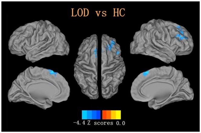
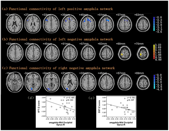
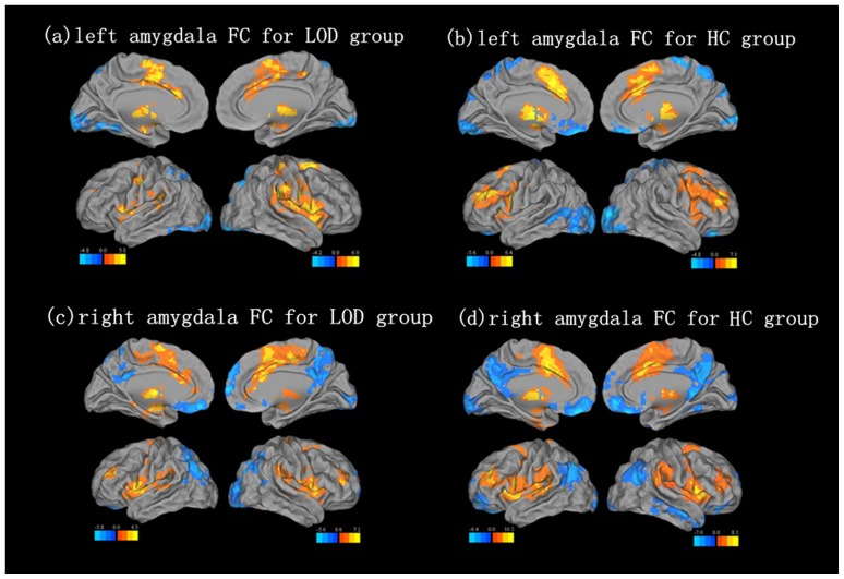
A total of twenty-two LOD patients and twenty-two matched healthy controls (HC) underwent neuropsychological tests and resting state functional magnetic resonance imaging (rs-fMRI). Regional homogeneity (ReHo) and FC with bilateral amygdala seeds were used to analyze blood oxygen level-dependent fMRI data between two groups. Compared with HC, LOD patients showed decreased ReHo in the right middle frontal gyrus and left superior frontal gyrus. In the LOD group, the left amygdala had decreased FC with the right middle frontal gyrus and the left superior frontal gyrus in the amygdala positive network, and it had increased FC with the right post-central gyrus in the amygdala negative network. However, significantly reduced FC with the right amygdala was observed in the right middle occipital gyrus in the amygdala negative network. Further correlative analyses revealed that decreased FC between the amygdala and the right middle occipital gyrus was negatively correlated with the verbal fluency test (VFT, r = -0.485, P = 0.022) and the digit span test (DST, r = -0.561, P = 0.007).
Our findings of reduced activity of the prefrontal gyrus and abnormal FC with the bilateral amygdala may be key markers of cognitive dysfunction in LOD patients.
重度抑郁症(MDD)与皮质-边缘回路功能下降有关,而皮质-边缘回路在 MDD 的发病机制中起着重要作用。MDD 患者的杏仁核等皮质-边缘回路也存在异常功能连接(FC)。然而,对于迟发性抑郁症(LOD)中连接变化的了解甚少,或者连接中断是否与 LOD 患者的认知障碍相关。
共纳入 22 名 LOD 患者和 22 名匹配的健康对照者(HC)进行神经心理学测试和静息态功能磁共振成像(rs-fMRI)检查。采用局部一致性(ReHo)和双侧杏仁核种子点 FC 分析两组间的血氧水平依赖 fMRI 数据。与 HC 相比,LOD 患者右侧额中回和左侧额上回的 ReHo 降低。在 LOD 组中,左侧杏仁核与右侧额中回和左侧额上回的 FC 在杏仁核正网络中降低,与右侧中央后回的 FC 在杏仁核负网络中增加。然而,杏仁核负网络中的右侧中枕叶与右侧杏仁核的 FC 显著降低。进一步的相关分析表明,杏仁核与右侧中枕叶之间的 FC 降低与言语流畅性测试(VFT,r=-0.485,P=0.022)和数字跨度测试(DST,r=-0.561,P=0.007)呈负相关。
我们发现额叶回活动减少和双侧杏仁核异常 FC 可能是 LOD 患者认知功能障碍的关键标志物。