• 文献检索
  • 文档翻译
  • 深度研究
  • 学术资讯
  • Suppr Zotero 插件Zotero 插件
  • 邀请有礼
  • 套餐&价格
  • 历史记录
应用&插件
Suppr Zotero 插件Zotero 插件浏览器插件Mac 客户端Windows 客户端微信小程序
定价
高级版会员购买积分包购买API积分包
服务
文献检索文档翻译深度研究API 文档MCP 服务
关于我们
关于 Suppr公司介绍联系我们用户协议隐私条款
关注我们

Suppr 超能文献

核心技术专利:CN118964589B侵权必究
粤ICP备2023148730 号-1Suppr @ 2026

文献检索

告别复杂PubMed语法,用中文像聊天一样搜索,搜遍4000万医学文献。AI智能推荐,让科研检索更轻松。

立即免费搜索

文件翻译

保留排版,准确专业,支持PDF/Word/PPT等文件格式,支持 12+语言互译。

免费翻译文档

深度研究

AI帮你快速写综述,25分钟生成高质量综述,智能提取关键信息,辅助科研写作。

立即免费体验

异位空气所显示的解剖学隔室及其连接。

The anatomical compartments and their connections as demonstrated by ectopic air.

机构信息

Department of Imagiology, Hospital-Escola da Universidade Fernando Pessoa, Gondomar, Porto, Portugal,

出版信息

Insights Imaging. 2013 Dec;4(6):759-72. doi: 10.1007/s13244-013-0278-0. Epub 2013 Sep 25.

DOI:10.1007/s13244-013-0278-0
PMID:24065628
原文链接:https://pmc.ncbi.nlm.nih.gov/articles/PMC3846937/
Abstract

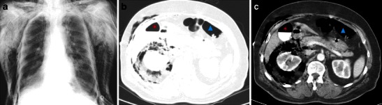
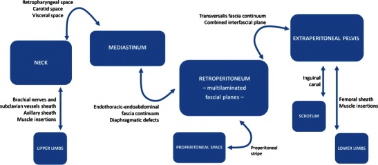
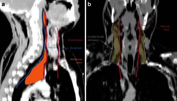
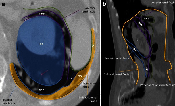
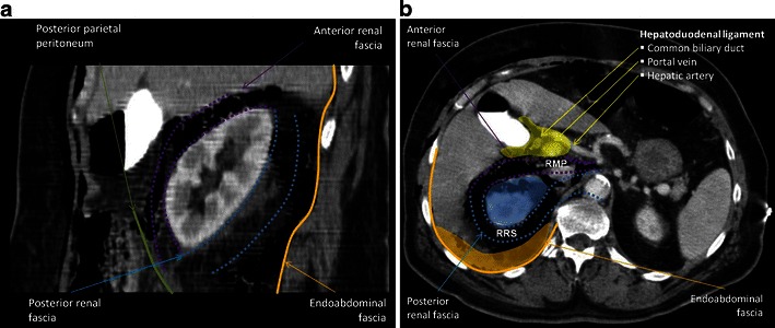
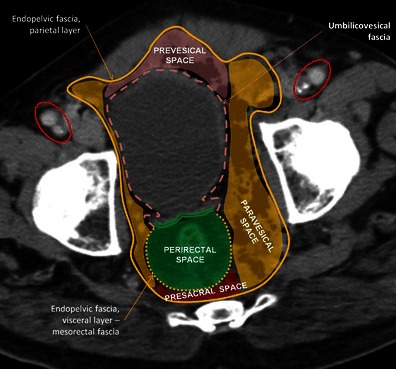
Air/gas outside the aero-digestive tract is abnormal; depending on its location, it is usually called emphysema, referring to trapped air/gas in tissues, or ectopic air/gas. It can be associated to a wide range of disorders, and although it usually is an innocuous condition, it should prompt a search for the underlying aetiology, since some of its causes impose an urgent treatment. In rare instances, it may itself represent a life-threatening condition, depending on the site involved and how quickly it evolves. Abnormal air/gas beyond viscera and serosal spaces, reaches its location following some anatomic boundaries, such as fascia, which may help search the source; however if the air pressure exceeds the strength of the tissues, or the time between the aggression and the imaging is too long, the air/gas is almost everywhere, which may hinder its cause. Good knowledge of the anatomic spaces and how they connect between them facilitates the quick detection of the cause. Teaching points • Ectopic air can be depicted on conventional radiographs; but CT is more sensitive and accurate • Visceral and retropharyngeal spaces directly communicate with mediastinum • Renal fascia is a single multilaminated structure, which contains potential space.

摘要

呼吸道外的气/气体会出现异常;根据其位置,通常被称为肺气肿,是指组织中滞留的气/气,或异位气/气。它可能与多种疾病相关,虽然通常是无害的,但应提示寻找潜在病因,因为一些病因需要紧急治疗。在极少数情况下,根据受累部位和演变速度,它本身可能是危及生命的情况。异常气/气超出内脏和浆膜间隙,沿某些解剖边界到达其位置,例如筋膜,这有助于寻找源头;但是如果气压超过组织强度,或者侵袭和成像之间的时间过长,气/气几乎无处不在,这可能会阻碍病因的确定。良好的解剖间隙知识以及它们之间的连接方式有助于快速发现病因。教学要点 • 异位气可在常规 X 线片上显示;但 CT 更敏感和准确 • 内脏和咽后间隙直接与纵隔相通 • 肾筋膜是一种单一的多层结构,包含潜在间隙。

https://cdn.ncbi.nlm.nih.gov/pmc/blobs/09f9/3846937/b74276bce9f3/13244_2013_278_Fig16_HTML.jpg
https://cdn.ncbi.nlm.nih.gov/pmc/blobs/09f9/3846937/0e94c67bf60c/13244_2013_278_Fig1_HTML.jpg
https://cdn.ncbi.nlm.nih.gov/pmc/blobs/09f9/3846937/d5c01711fa2c/13244_2013_278_Fig2_HTML.jpg
https://cdn.ncbi.nlm.nih.gov/pmc/blobs/09f9/3846937/facc01b8b2ac/13244_2013_278_Fig3_HTML.jpg
https://cdn.ncbi.nlm.nih.gov/pmc/blobs/09f9/3846937/d9bc1215e0b3/13244_2013_278_Fig4_HTML.jpg
https://cdn.ncbi.nlm.nih.gov/pmc/blobs/09f9/3846937/20b179977201/13244_2013_278_Fig5_HTML.jpg
https://cdn.ncbi.nlm.nih.gov/pmc/blobs/09f9/3846937/095ea976e2fa/13244_2013_278_Fig6_HTML.jpg
https://cdn.ncbi.nlm.nih.gov/pmc/blobs/09f9/3846937/c537cd0cdc63/13244_2013_278_Fig7_HTML.jpg
https://cdn.ncbi.nlm.nih.gov/pmc/blobs/09f9/3846937/db03e17b958f/13244_2013_278_Fig8_HTML.jpg
https://cdn.ncbi.nlm.nih.gov/pmc/blobs/09f9/3846937/99fb5ca76d2a/13244_2013_278_Fig9_HTML.jpg
https://cdn.ncbi.nlm.nih.gov/pmc/blobs/09f9/3846937/d08387bcd1fa/13244_2013_278_Fig10_HTML.jpg
https://cdn.ncbi.nlm.nih.gov/pmc/blobs/09f9/3846937/e8b1d5c7eb0c/13244_2013_278_Fig11_HTML.jpg
https://cdn.ncbi.nlm.nih.gov/pmc/blobs/09f9/3846937/7be3cc718019/13244_2013_278_Fig12_HTML.jpg
https://cdn.ncbi.nlm.nih.gov/pmc/blobs/09f9/3846937/4a85b5013c6d/13244_2013_278_Fig13_HTML.jpg
https://cdn.ncbi.nlm.nih.gov/pmc/blobs/09f9/3846937/ef85b9acee99/13244_2013_278_Fig14_HTML.jpg
https://cdn.ncbi.nlm.nih.gov/pmc/blobs/09f9/3846937/e657755dd899/13244_2013_278_Fig15_HTML.jpg
https://cdn.ncbi.nlm.nih.gov/pmc/blobs/09f9/3846937/b74276bce9f3/13244_2013_278_Fig16_HTML.jpg

相似文献

1
The anatomical compartments and their connections as demonstrated by ectopic air.异位空气所显示的解剖学隔室及其连接。
Insights Imaging. 2013 Dec;4(6):759-72. doi: 10.1007/s13244-013-0278-0. Epub 2013 Sep 25.
2
Imaging Assessment of Ectopic Gas Collections.异位气体积聚的影像学评估。
Radiographics. 2020 Sep-Oct;40(5):1318-1338. doi: 10.1148/rg.2020200028. Epub 2020 Aug 14.
3
Thoracic abnormal air collections in patients in the intensive care unit: radiograph findings correlated with CT.重症监护病房患者的胸部异常气体积聚:X线片表现与CT的相关性
Insights Imaging. 2020 Mar 12;11(1):35. doi: 10.1186/s13244-020-0838-z.
4
Cervicofacial and mediastinal emphysema after dental extraction.拔牙后颈面部及纵隔气肿
Dent Med Probl. 2019 Apr-Jun;56(2):203-207. doi: 10.17219/dmp/108615.
5
DESCENDING NECROTIZING MEDIASTINITIS SECONDARY TO RETROPHARYNGEAL ABSCESS.继发于咽后脓肿的下行性坏死性纵隔炎
Acta Clin Croat. 2015 Dec;54(4):541-6.
6
[The posterior interparieto-peritoneal or retroperitoneal spaces. 2: Pathological x-ray computed tomographic image].
J Radiol. 1991 Apr;72(4):205-27.
7
Scary gas: pathways in the axial body for soft tissue gas dissection (part I).可怕的气体:轴向身体软组织气体剥离的路径(第一部分)
Emerg Radiol. 2017 Oct;24(5):569-576. doi: 10.1007/s10140-017-1489-3. Epub 2017 Mar 1.
8
Cervicofacial emphysema after dental treatment with emphasis on the anatomy of the cervical fascia.
J Craniofac Surg. 2012 Nov;23(6):e544-8. doi: 10.1097/SCS.0b013e31825aef02.
9
Scary gas: a spectrum of soft tissue gas encountered in the axial body (part II).可怕的气体:轴向身体中遇到的软组织气体谱(第二部分)
Emerg Radiol. 2017 Aug;24(4):401-409. doi: 10.1007/s10140-017-1491-9. Epub 2017 Mar 2.
10
[Subcutaneous emphysema after septoplasty: a case report].[鼻中隔成形术后皮下气肿:一例报告]
Kulak Burun Bogaz Ihtis Derg. 2016;26(1):51-4. doi: 10.5606/kbbihtisas.2016.92593.

引用本文的文献

1
Successful management of severe anaerobic infection inducing mediastinal and subcutaneous emphysema, septic shock, and ARDS: a case report highlighting VV-ECMO and NGS-guided therapy.成功治疗导致纵隔和皮下气肿、感染性休克及急性呼吸窘迫综合征的严重厌氧感染:一例强调静脉-静脉体外膜肺氧合及二代测序引导治疗的病例报告
Front Med (Lausanne). 2025 Mar 17;12:1568410. doi: 10.3389/fmed.2025.1568410. eCollection 2025.
2
Incidence, outcomes, and risk factors of postlaparoscopic subcutaneous emphysema: a historical cohort study.腹腔镜术后皮下气肿的发生率、结局及危险因素:一项历史性队列研究。
Can J Anaesth. 2025 Jan;72(1):152-161. doi: 10.1007/s12630-024-02859-2. Epub 2024 Oct 17.
3

本文引用的文献

1
Crossing the barrier: when the diaphragm is not a limit.穿越障碍:当横膈膜不再是限制。
AJR Am J Roentgenol. 2013 Jan;200(1):W62-70. doi: 10.2214/AJR.11.8264.
2
Imaging anatomy of deep neck spaces.颈部深部间隙的影像解剖
Otolaryngol Clin North Am. 2012 Dec;45(6):1203-21. doi: 10.1016/j.otc.2012.08.001.
3
Fascia: a morphological description and classification system based on a literature review.筋膜:基于文献综述的形态学描述与分类系统
Pneumothorax, Pneumomediastinum, and Cervical and Facial Massive Emphysema Secondary to Colonoscopy: A Rare Complication of Colonoscopy.
结肠镜检查继发气胸、纵隔气肿以及颈部和面部大面积气肿:结肠镜检查的一种罕见并发症
Case Rep Gastrointest Med. 2024 Jun 25;2024:1140099. doi: 10.1155/2024/1140099. eCollection 2024.
4
Manual Therapy of Dysphagia in a Patient with Amyotrophic Lateral Sclerosis: A Case Report.肌萎缩侧索硬化症患者吞咽障碍的手法治疗:病例报告。
Medicina (Kaunas). 2024 May 22;60(6):845. doi: 10.3390/medicina60060845.
5
Dirty Mass sign of abdominal imaging: A case of silent pneumoperitoneum.腹部影像学的脏块征:一例隐匿性气腹病例。
Radiol Case Rep. 2024 Feb 22;19(5):1839-1842. doi: 10.1016/j.radcr.2024.02.012. eCollection 2024 May.
6
Retroperitoneal anatomy with the aid of pathologic fluid: An imaging pictorial review.借助病理性液体的腹膜后解剖:影像学图谱综述
J Clin Imaging Sci. 2023 Dec 13;13:36. doi: 10.25259/JCIS_79_2023. eCollection 2023.
7
Air Will Find a Way: A Case Report and Literature Review on Tracheostomy-Induced Ectopic Air.空气总会找到出路:一例气管切开术致异位空气的病例报告及文献综述
Cureus. 2023 Jul 25;15(7):e42446. doi: 10.7759/cureus.42446. eCollection 2023 Jul.
8
Case series: Pleural effusion caused by urinary ultrafiltrate in two cats without evidence of urinary obstruction, trauma, or simultaneous perinephric pseudocysts.
Front Vet Sci. 2022 Nov 21;9:1038278. doi: 10.3389/fvets.2022.1038278. eCollection 2022.
9
The Mediastinal Visceral Space: The Central Pathway for the Spread of Mediastinal Disease.纵隔内脏间隙:纵隔疾病扩散的中心途径。
Indian J Radiol Imaging. 2022 Aug 17;32(3):365-371. doi: 10.1055/s-0042-1756221. eCollection 2022 Sep.
10
Imaging evaluation of uterine perforation and rupture.子宫穿孔和破裂的影像学评估。
Abdom Radiol (NY). 2021 Oct;46(10):4946-4966. doi: 10.1007/s00261-021-03171-z. Epub 2021 Jun 15.
J Can Chiropr Assoc. 2012 Sep;56(3):179-91.
4
Peritoneal and retroperitoneal anatomy and its relevance for cross-sectional imaging.腹膜和腹膜后解剖及其与横断影像学的相关性。
Radiographics. 2012 Mar-Apr;32(2):437-51. doi: 10.1148/rg.322115032.
5
Maneuvers to decrease laparoscopy-induced shoulder and upper abdominal pain: a randomized controlled study.减轻腹腔镜手术引起的肩部和上腹部疼痛的手法:一项随机对照研究。
Arch Surg. 2011 Dec;146(12):1360-6. doi: 10.1001/archsurg.2011.597.
6
Pathways of extrapelvic spread of pelvic disease: imaging findings.盆腔疾病盆外蔓延途径:影像学表现。
Radiographics. 2011 Jan-Feb;31(1):117-33. doi: 10.1148/rg.311105050.
7
Successful Nonsurgical Treatment of Pneumomediastinum, Pneumothorax, Pneumoperitoneum, Pneumoretroperitoneum, and Subcutaneous Emphysema following ERCP.ERCP 后并发纵隔气肿、气胸、气腹、腹膜后积气和皮下气肿的成功非手术治疗。
Gastroenterol Res Pract. 2010;2010:289135. doi: 10.1155/2010/289135. Epub 2010 Jun 14.
8
[Massive subcutaneous emphysema--management using subcutaneous drains].[巨大皮下气肿——使用皮下引流管的处理方法]
Rev Port Pneumol. 2010 Mar-Apr;16(2):321-9. doi: 10.1016/s0873-2159(15)30030-1.
9
Radiologic and anatomic study of the extraperitoneal space associated with the rectum.直肠周围腹膜外间隙的放射学与解剖学研究。
AJR Am J Roentgenol. 2010 Mar;194(3):642-52. doi: 10.2214/AJR.09.3003.
10
Comprehensive reviews of the interfascial plane of the retroperitoneum: normal anatomy and pathologic entities.腹膜后间隙筋膜平面的综合综述:正常解剖与病理实体
Emerg Radiol. 2010 Jan;17(1):3-11. doi: 10.1007/s10140-009-0809-7. Epub 2009 Apr 28.