Department of Surgery, University of Texas Health Science Center at Houston, Houston, Texas, United States of America.
PLoS One. 2013 Sep 20;8(9):e76790. doi: 10.1371/journal.pone.0076790. eCollection 2013.
The role of extracellular signal-regulated protein kinase (ERK) in intestinal ischemia/reperfusion (I/R) injury has not been well investigated. The aim of the current study was to examine the effect of inhibition of the ERK pathway in an in vitro and in vivo model of intestinal I/R injury.
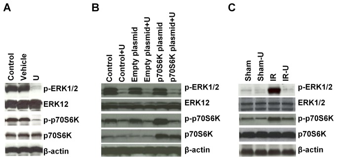
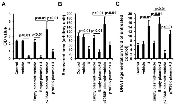
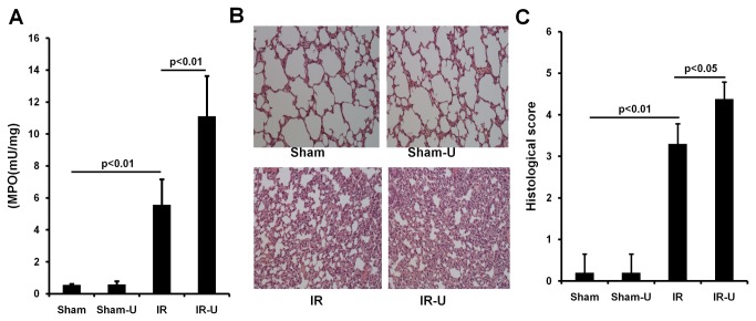
ERK1/2 activity was inhibited using the specific inhibitor, U0126, in intestinal epithelial cells under hypoxia/reoxygenation conditions and in mice subjected to 1 hour of intestinal ischemia followed by 6 hours reperfusion. In vitro, cell proliferation was assessed by MTT (3-(4,5-dimethylthiazol-2-yl)-2,5-diphenyl tetrazolium bromide) assay, apoptosis by DNA fragmentation, and migration using an in vitro model of intestinal wound healing. Cells were also transfected with a p70S6K plasmid and the effects of overexpression similarly analyzed. In vivo, the effects of U0126 on intestinal cell proliferation and apoptosis, intestinal permeability, lung and intestinal neutrophil infiltration and injury, and plasma cytokine levels were measured. Survival was also assessed after U0126. Activity of p70S6 kinase (p70S6K) was measured by Western blot.
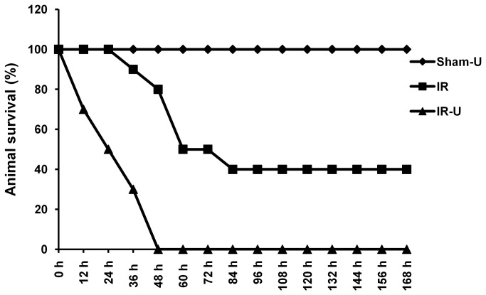
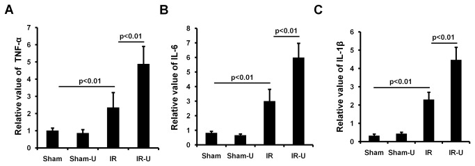
In vitro, inhibition of ERK1/2 by U0126 significantly decreased cell proliferation and migration but enhanced cell apoptosis. Overexpression of p70S6K promoted cell proliferation and decreased cell apoptosis. In vivo, U0126 significantly increased cell apoptosis and decreased cell proliferation in the intestine, increased intestinal permeability, intestinal and lung neutrophil infiltration, and injury, as well as systemic pro-inflammatory cytokines, TNF-α, IL-6 and IL-1β. Mortality was also significantly increased by U0126. Inhibition of ERK1/2 by U0126 also abolished activity of p70S6K both in vitro and in vivo models.
Pharmacologic inhibition of ERK1/2 by U0126 worsens intestinal IR injury. The detrimental effects are mediated, at least in part, by inhibition of p70S6K, the major effector of mammalian target of rapamycin pathway.
细胞外信号调节激酶(ERK)在肠缺血/再灌注(I/R)损伤中的作用尚未得到充分研究。本研究旨在研究在肠 I/R 损伤的体外和体内模型中抑制 ERK 通路的效果。
在缺氧/复氧条件下的肠上皮细胞中和在经历 1 小时肠缺血后再灌注 6 小时的小鼠中,使用特定的 ERK1/2 抑制剂 U0126 抑制 ERK1/2 活性。在体外,通过 MTT(3-(4,5-二甲基噻唑-2-基)-2,5-二苯基四唑溴盐)测定法评估细胞增殖,通过 DNA 片段化评估细胞凋亡,通过肠伤口愈合的体外模型评估细胞迁移。还转染了 p70S6K 质粒,并同样分析了过表达的效果。在体内,测量了 U0126 对肠细胞增殖和凋亡、肠通透性、肺和肠中性粒细胞浸润和损伤以及血浆细胞因子水平的影响。还评估了 U0126 后的存活率。通过 Western blot 测定 p70S6 激酶(p70S6K)的活性。
在体外,U0126 抑制 ERK1/2 显著降低了细胞增殖和迁移,但增强了细胞凋亡。p70S6K 的过表达促进了细胞增殖并减少了细胞凋亡。在体内,U0126 显著增加了肠内细胞凋亡并减少了细胞增殖,增加了肠通透性,肠和肺中性粒细胞浸润和损伤,以及全身促炎细胞因子 TNF-α、IL-6 和 IL-1β。U0126 还显著增加了死亡率。U0126 还在体外和体内模型中抑制了 ERK1/2 的活性。
U0126 通过药理学抑制 ERK1/2 加重了肠 I/R 损伤。有害作用至少部分是通过抑制 p70S6K 介导的,p70S6K 是哺乳动物雷帕霉素靶蛋白途径的主要效应物。