Department of Neurology, Washington University, St. Louis, MO, USA.
Neuroimage Clin. 2013 Jun 25;2:862-72. doi: 10.1016/j.nicl.2013.06.011. eCollection 2013.
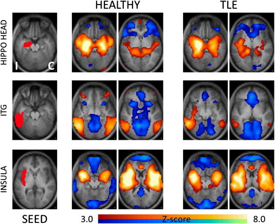
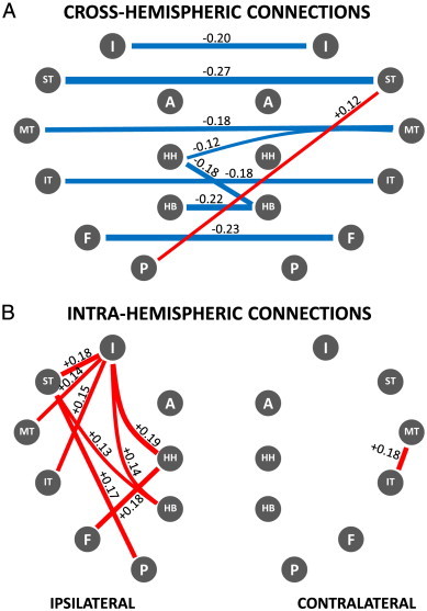
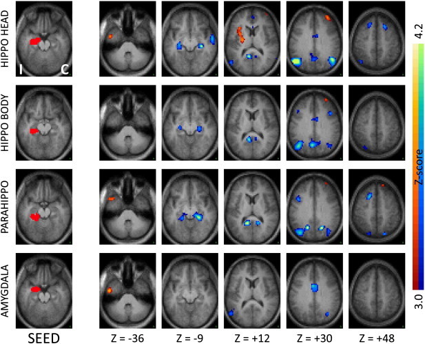
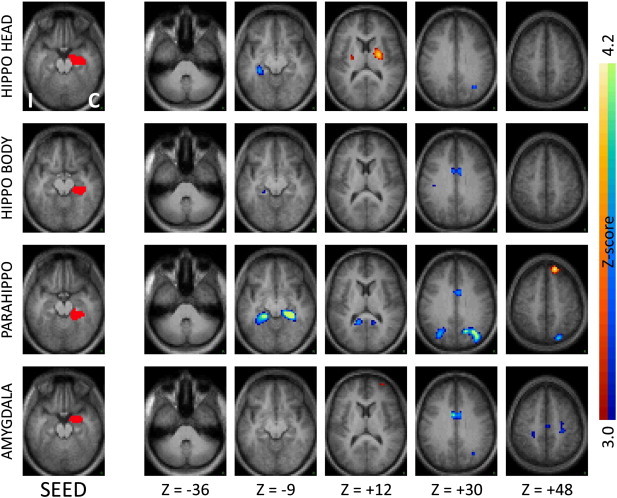
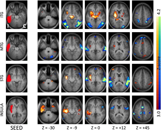
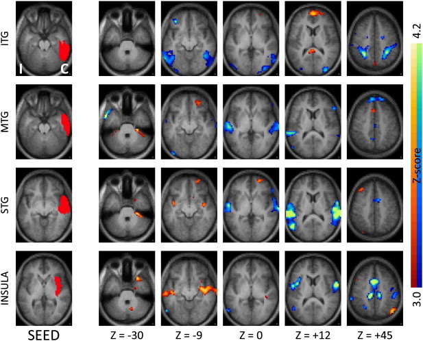
How epilepsy affects brain functional networks remains poorly understood. Here we investigated resting state functional connectivity of the temporal region in temporal lobe epilepsy. Thirty-two patients with unilateral temporal lobe epilepsy underwent resting state blood-oxygenation level dependent functional magnetic resonance imaging. We defined regions of interest a priori focusing on structures involved, either structurally or metabolically, in temporal lobe epilepsy. These structures were identified in each patient based on their individual anatomy. Our principal findings are decreased local and inter-hemispheric functional connectivity and increased intra-hemispheric functional connectivity ipsilateral to the seizure focus compared to normal controls. Specifically, several regions in the affected temporal lobe showed increased functional coupling with the ipsilateral insula and immediately neighboring subcortical regions. Additionally there was significantly decreased functional connectivity between regions in the affected temporal lobe and their contralateral homologous counterparts. Intriguingly, decreased local and inter-hemispheric connectivity was not limited or even maximal for the hippocampus or medial temporal region, which is the typical seizure onset region. Rather it also involved several regions in temporal neo-cortex, while also retaining specificity, with neighboring regions such as the amygdala remaining unaffected. These findings support a view of temporal lobe epilepsy as a disease of a complex functional network, with alterations that extend well beyond the seizure onset area, and the specificity of the observed connectivity changes suggests the possibility of a functional imaging biomarker for temporal lobe epilepsy.
癫痫如何影响大脑功能网络仍知之甚少。在这里,我们研究了颞叶癫痫的颞叶区域的静息状态功能连接。32 例单侧颞叶癫痫患者接受了静息状态血氧水平依赖功能磁共振成像。我们以前瞻性的方式定义了感兴趣的区域,重点关注颞叶癫痫中结构或代谢上涉及的结构。这些结构是根据每个患者的个体解剖结构在每个患者中确定的。我们的主要发现是与正常对照组相比,癫痫灶同侧的局部和半球间功能连接减少,半球内功能连接增加。具体来说,受影响的颞叶中的几个区域与同侧岛叶和紧邻的皮质下区域显示出增加的功能耦合。此外,受影响的颞叶区域与其对侧同源区域之间的功能连接显著降低。有趣的是,局部和半球间连接的减少不仅限于甚至对于海马体或内侧颞叶区域(即典型的癫痫发作起始区域)最大。相反,它还涉及颞叶新皮质中的几个区域,同时保持特异性,相邻区域如杏仁核不受影响。这些发现支持了这样一种观点,即颞叶癫痫是一种复杂功能网络疾病,其改变远远超出了癫痫发作起始区域,并且观察到的连接变化的特异性表明颞叶癫痫可能存在功能成像生物标志物。