*Department of Pathology, Division of Women's and Perinatal Pathology §Department of Obstetrics and Gynecology and Reproductive Biology, Brigham and Women's Hospital, Boston, MA ‡Department of Pathology, Cedar Sinai Medical Center, Los Angeles, CA †Department of Pathology, GIGA-Cancer, University of Liege, Liege, Belgium ∥Genome Institute of Singapore, ASTAR The Jackson Laboratory for Genomic Medicine, CT ¶The Jackson Laboratory for Genomic Medicine, The University of Connecticut School of Medicine, CT.
Am J Surg Pathol. 2013 Sep;37(9):1311-8. doi: 10.1097/PAS.0b013e3182989ee2.
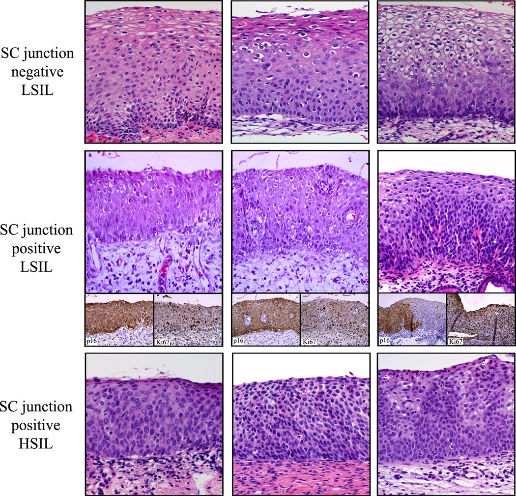
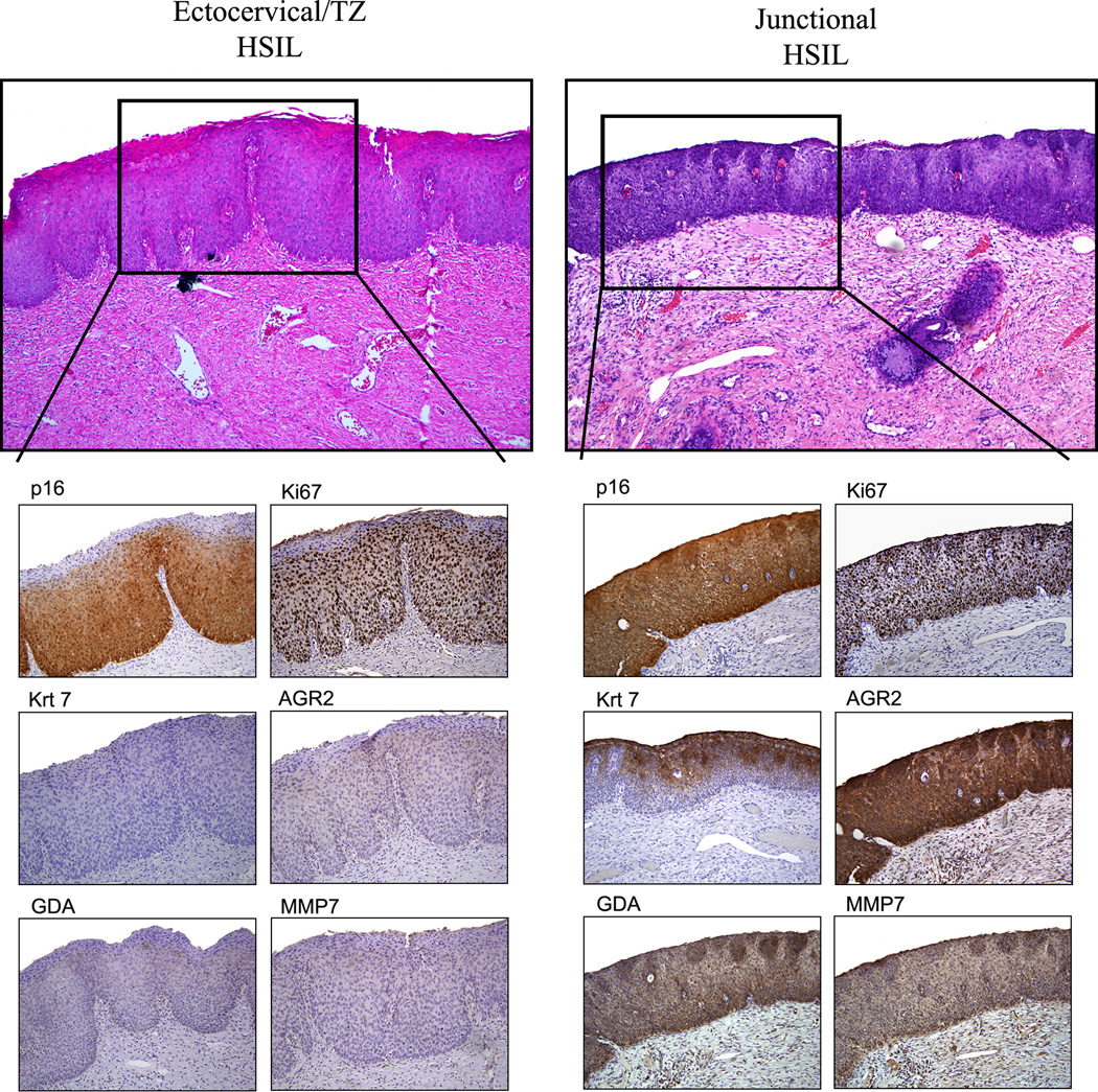
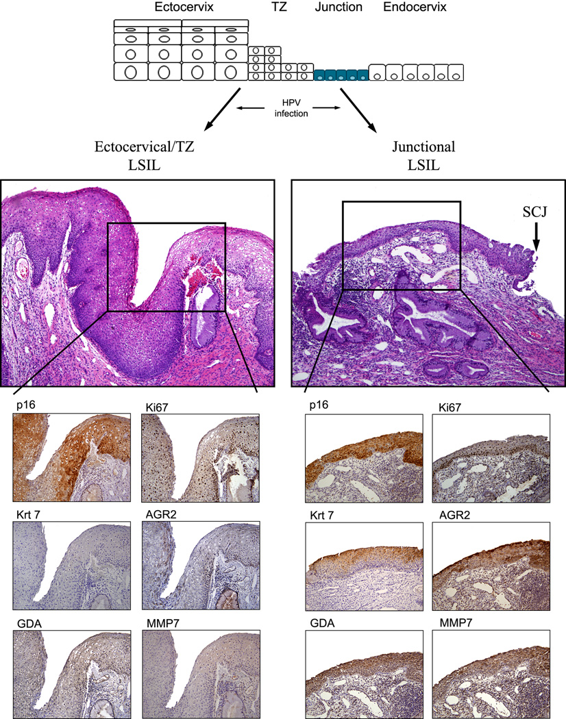
Low-grade cervical squamous abnormalities (low-grade squamous intraepithelial lesions [LSIL, CIN1]) can be confused with or followed by high-grade (HSIL, CIN2/3) lesions, expending considerable resources. Recently, a cell of origin for cervical neoplasia was proposed in the squamocolumnar junction (SCJ); HSILs are almost always SCJ, but LSILs include SCJ and SCJ subsets. Abnormal cervical biopsies from 214 patients were classified by 2 experienced pathologists (panel) as LSIL or HSIL using published criteria. SILs were scored SCJ and SCJ using SCJ-specific antibodies (keratin7, AGR2, MMP7, and GDA). Assessments of interobserver agreement, p16 staining pattern, proliferative index, and outcome were compared. The original diagnostician agreed with the panel diagnosis of HSIL and SCJ LSIL in all cases (100%). However, for SCJ LSIL, panelists disagreed with each other by 15% and with the original diagnostician by 46.2%. Comparing SCJ and SCJ LSILs, 60.2% and 94.9% were p16 positive, 23% and 74.4% showed strong (full-thickness) p16 staining, and 0/54 (0%) and 8/33 (24.2%) with follow-up had an HSIL outcome, respectively. Some SCJ LSILs are more likely to both generate diagnostic disagreement and be associated with HSIL. Conversely, SCJ LSILs generate little observer disagreement and, when followed, have a very low risk of HSIL outcome. Thus, SCJ biomarkers in conjunction with histology may segregate LSILs with very low risk of HSIL outcome and conceivably could be used as a management tool to reduce excess allocation of resources to the follow-up of these lesions.
低度宫颈鳞状上皮内病变(低度鳞状上皮内病变 [LSIL,CIN1])可能与高度病变(HSIL,CIN2/3)混淆或随后发生,耗费大量资源。最近,宫颈肿瘤的细胞起源被提出在鳞柱状交界区(SCJ);HSIL 几乎总是 SCJ,但 LSIL 包括 SCJ 和 SCJ 亚群。2 名经验丰富的病理学家(小组)使用发表的标准,将 214 名患者的异常宫颈活检分为 LSIL 或 HSIL。使用 SCJ 特异性抗体(角蛋白 7、AGR2、MMP7 和 GDA)对 SIL 进行 SCJ 和 SCJ 评分。比较了观察者间的一致性、p16 染色模式、增殖指数和结果。原始诊断医生与小组诊断的 HSIL 和 SCJ LSIL 完全一致(100%)。然而,对于 SCJ LSIL,小组成员之间的意见分歧为 15%,与原始诊断医生的意见分歧为 46.2%。比较 SCJ 和 SCJ LSIL,60.2%和 94.9%为 p16 阳性,23%和 74.4%显示强(全层)p16 染色,54 个中没有(0%)和 33 个中有 8 个(24.2%)有 HSIL 结果,分别。一些 SCJ LSIL 更有可能产生诊断分歧,并与 HSIL 相关。相反,SCJ LSIL 产生的观察者分歧较小,并且在随访时 HSIL 结果的风险非常低。因此,SCJ 生物标志物与组织学相结合可能会将 HSIL 结果风险非常低的 LSIL 分开,并且可以设想将其用作管理工具,以减少对这些病变随访的过度资源分配。