• 文献检索
  • 文档翻译
  • 深度研究
  • 学术资讯
  • Suppr Zotero 插件Zotero 插件
  • 邀请有礼
  • 套餐&价格
  • 历史记录
应用&插件
Suppr Zotero 插件Zotero 插件浏览器插件Mac 客户端Windows 客户端微信小程序
定价
高级版会员购买积分包购买API积分包
服务
文献检索文档翻译深度研究API 文档MCP 服务
关于我们
关于 Suppr公司介绍联系我们用户协议隐私条款
关注我们

Suppr 超能文献

核心技术专利:CN118964589B侵权必究
粤ICP备2023148730 号-1Suppr @ 2026

文献检索

告别复杂PubMed语法,用中文像聊天一样搜索,搜遍4000万医学文献。AI智能推荐,让科研检索更轻松。

立即免费搜索

文件翻译

保留排版,准确专业,支持PDF/Word/PPT等文件格式,支持 12+语言互译。

免费翻译文档

深度研究

AI帮你快速写综述,25分钟生成高质量综述,智能提取关键信息,辅助科研写作。

立即免费体验

从微秒全原子分子动力学模拟中得出的脂质受体 S1P₁激活方案。

Lipid receptor S1P₁ activation scheme concluded from microsecond all-atom molecular dynamics simulations.

机构信息

International Institute of Molecular and Cell Biology in Warsaw, Warsaw, Poland ; Laboratory of Physical Chemistry of Polymers and Membranes, École Polytechnique Fédérale de Lausanne, SB ISIC LCPPM, Lausanne, Switzerland ; Nencki Institute of Experimental Biology, Polish Academy of Sciences, Warsaw, Poland.

出版信息

PLoS Comput Biol. 2013;9(10):e1003261. doi: 10.1371/journal.pcbi.1003261. Epub 2013 Oct 3.

DOI:10.1371/journal.pcbi.1003261
PMID:24098103
原文链接:https://pmc.ncbi.nlm.nih.gov/articles/PMC3789783/
Abstract

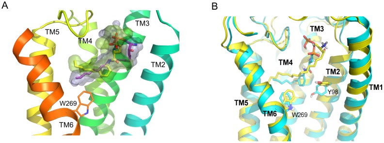
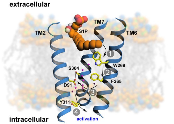
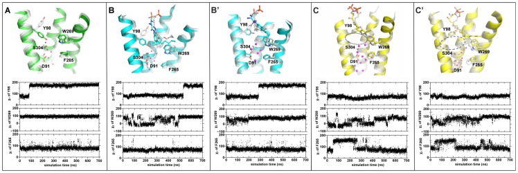
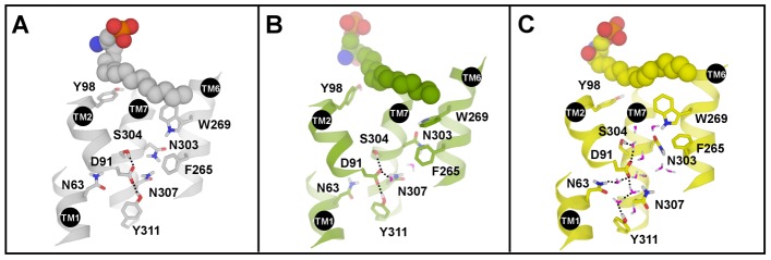
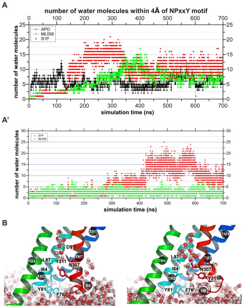
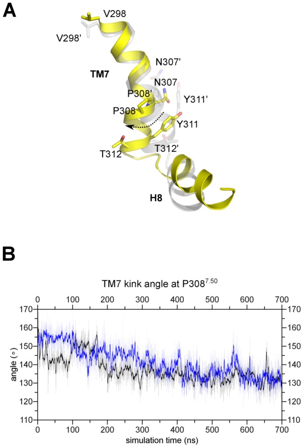
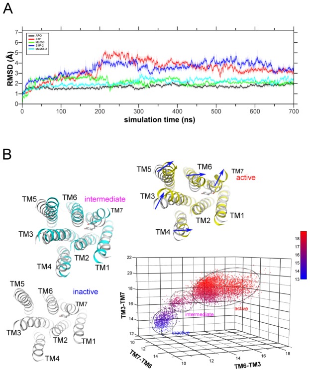
Sphingosine 1-phosphate (S1P) is a lysophospholipid mediator which activates G protein-coupled sphingosine 1-phosphate receptors and thus evokes a variety of cell and tissue responses including lymphocyte trafficking, endothelial development, integrity, and maturation. We performed five all-atom 700 ns molecular dynamics simulations of the sphingosine 1-phosphate receptor 1 (S1P₁) based on recently released crystal structure of that receptor with an antagonist. We found that the initial movements of amino acid residues occurred in the area of highly conserved W269⁶·⁴⁸ in TM6 which is close to the ligand binding location. Those residues located in the central part of the receptor and adjacent to kinks of TM helices comprise of a transmission switch. Side chains movements of those residues were coupled to the movements of water molecules inside the receptor which helped in the gradual opening of intracellular part of the receptor. The most stable parts of the protein were helices TM1 and TM2, while the largest movement was observed for TM7, possibly due to the short intracellular part starting with a helix kink at P⁷·⁵⁰, which might be the first helix to move at the intracellular side. We show for the first time the detailed view of the concerted action of the transmission switch and Trp (W⁶·⁴⁸) rotamer toggle switch leading to redirection of water molecules flow in the central part of the receptor. That event is a prerequisite for subsequent changes in intracellular part of the receptor involving water influx and opening of the receptor structure.

摘要

鞘氨醇 1-磷酸(S1P)是一种溶血磷脂介质,可激活 G 蛋白偶联鞘氨醇 1-磷酸受体,从而引发多种细胞和组织反应,包括淋巴细胞迁移、内皮细胞发育、完整性和成熟。我们对基于最近发布的该受体与拮抗剂的晶体结构的 1 型鞘氨醇 1-磷酸受体(S1P₁)进行了五次全原子 700 ns 分子动力学模拟。我们发现,氨基酸残基的初始运动发生在 TM6 中高度保守的 W269⁶·⁴⁸区域,该区域靠近配体结合位置。这些位于受体中央部分且靠近 TM 螺旋拐点的残基构成了一个传递开关。这些残基的侧链运动与受体内部水分子的运动耦合在一起,有助于受体细胞内部分的逐渐打开。蛋白质最稳定的部分是 TM1 和 TM2 螺旋,而 TM7 发生的运动最大,这可能是由于起始于 P⁷·⁵⁰处螺旋拐点的短细胞内部分,这可能是第一个在细胞内侧移动的螺旋。我们首次展示了传递开关和色氨酸(W⁶·⁴⁸)旋转体切换协同作用的详细视图,导致水分子在受体中央部分的流向发生重定向。该事件是受体细胞内部分随后变化的先决条件,涉及水分子内流和受体结构的打开。

https://cdn.ncbi.nlm.nih.gov/pmc/blobs/d4c4/3789783/37b43f0b3a06/pcbi.1003261.g007.jpg
https://cdn.ncbi.nlm.nih.gov/pmc/blobs/d4c4/3789783/2a35b01ca8e5/pcbi.1003261.g001.jpg
https://cdn.ncbi.nlm.nih.gov/pmc/blobs/d4c4/3789783/d7a254d25a87/pcbi.1003261.g002.jpg
https://cdn.ncbi.nlm.nih.gov/pmc/blobs/d4c4/3789783/068f1f09731f/pcbi.1003261.g003.jpg
https://cdn.ncbi.nlm.nih.gov/pmc/blobs/d4c4/3789783/c937bbceff55/pcbi.1003261.g004.jpg
https://cdn.ncbi.nlm.nih.gov/pmc/blobs/d4c4/3789783/cc7ffdeb59e4/pcbi.1003261.g005.jpg
https://cdn.ncbi.nlm.nih.gov/pmc/blobs/d4c4/3789783/e5c18b5874eb/pcbi.1003261.g006.jpg
https://cdn.ncbi.nlm.nih.gov/pmc/blobs/d4c4/3789783/37b43f0b3a06/pcbi.1003261.g007.jpg

相似文献

1
Lipid receptor S1P₁ activation scheme concluded from microsecond all-atom molecular dynamics simulations.从微秒全原子分子动力学模拟中得出的脂质受体 S1P₁激活方案。
PLoS Comput Biol. 2013;9(10):e1003261. doi: 10.1371/journal.pcbi.1003261. Epub 2013 Oct 3.
2
Ligand chain length drives activation of lipid G protein-coupled receptors.配体链长驱动脂类 G 蛋白偶联受体的激活。
Sci Rep. 2017 May 17;7(1):2020. doi: 10.1038/s41598-017-02104-5.
3
Activation mechanisms of the first sphingosine-1-phosphate receptor.首个1-磷酸鞘氨醇受体的激活机制
Protein Sci. 2017 Jun;26(6):1150-1160. doi: 10.1002/pro.3165. Epub 2017 Apr 12.
4
Chemical and genetic tools to explore S1P biology.探索 S1P 生物学的化学和遗传工具。
Curr Top Microbiol Immunol. 2014;378:55-83. doi: 10.1007/978-3-319-05879-5_3.
5
Sphingosine 1-phosphate analogue recognition and selectivity at S1P4 within the endothelial differentiation gene family of receptors.在内皮分化基因受体家族中,鞘氨醇-1-磷酸类似物对S1P4的识别及选择性
Biochem J. 2005 Jul 1;389(Pt 1):187-95. doi: 10.1042/BJ20050046.
6
Binding Characteristics of Sphingosine-1-Phosphate to ApoM hints to Assisted Release Mechanism via the ApoM Calyx-Opening.鞘氨醇-1-磷酸与载脂蛋白M的结合特性提示通过载脂蛋白M萼片开放的辅助释放机制。
Sci Rep. 2016 Aug 1;6:30655. doi: 10.1038/srep30655.
7
Molecular Mechanism of S1P Binding and Activation of the S1P Receptor.S1P 受体结合和激活的分子机制。
J Chem Inf Model. 2019 Oct 28;59(10):4402-4412. doi: 10.1021/acs.jcim.9b00642. Epub 2019 Oct 15.
8
Structure of the first sphingosine 1-phosphate receptor.首个神经酰胺 1-磷酸受体结构。
Sci Signal. 2012 May 22;5(225):pe23. doi: 10.1126/scisignal.2003160.
9
A single amino acid determines preference between phospholipids and reveals length restriction for activation of the S1P4 receptor.单个氨基酸决定了对磷脂的偏好,并揭示了S1P4受体激活的长度限制。
BMC Biochem. 2004 Aug 6;5:12. doi: 10.1186/1471-2091-5-12.
10
Identification of the hydrophobic ligand binding pocket of the S1P1 receptor.1-磷酸鞘氨醇受体1(S1P1)疏水配体结合口袋的鉴定。
J Biol Chem. 2007 Jan 26;282(4):2374-85. doi: 10.1074/jbc.M609648200. Epub 2006 Nov 18.

引用本文的文献

1
Identification of potential immunomodulators from Pulsatilla decoction that act on therapeutic targets for ulcerative colitis based on pharmacological activity, absorbed ingredients, and in-silico molecular docking.基于药理活性、吸收成分和计算机模拟分子对接,从白头翁汤中鉴定作用于溃疡性结肠炎治疗靶点的潜在免疫调节剂。
Chin Med. 2022 Nov 24;17(1):132. doi: 10.1186/s13020-022-00684-7.
2
Structural basis for receptor selectivity and inverse agonism in S1P receptors.S1P 受体的受体选择性和反向激动作用的结构基础。
Nat Commun. 2022 Aug 12;13(1):4736. doi: 10.1038/s41467-022-32447-1.
3
Helix 8 in chemotactic receptors of the complement system.

本文引用的文献

1
Prediction of Absolute Solvation Free Energies using Molecular Dynamics Free Energy Perturbation and the OPLS Force Field.使用分子动力学自由能微扰和OPLS力场预测绝对溶剂化自由能
J Chem Theory Comput. 2010 May 11;6(5):1509-19. doi: 10.1021/ct900587b. Epub 2010 Apr 14.
2
A sphingosine 1-phosphate receptor 2 selective allosteric agonist.一种鞘氨醇 1-磷酸受体 2 选择性别构激动剂。
Bioorg Med Chem. 2013 Sep 1;21(17):5373-82. doi: 10.1016/j.bmc.2013.06.012. Epub 2013 Jun 15.
3
The role of water in activation mechanism of human N-formyl peptide receptor 1 (FPR1) based on molecular dynamics simulations.
螺旋 8 在补体系统趋化受体中的作用。
PLoS Comput Biol. 2022 Jul 21;18(7):e1009994. doi: 10.1371/journal.pcbi.1009994. eCollection 2022 Jul.
4
Recent Advances in Structure, Function, and Pharmacology of Class A Lipid GPCRs: Opportunities and Challenges for Drug Discovery.A类脂质G蛋白偶联受体的结构、功能与药理学研究进展:药物研发的机遇与挑战
Pharmaceuticals (Basel). 2021 Dec 22;15(1):12. doi: 10.3390/ph15010012.
5
Docking Studies of Fingolimod and S1P Agonists.芬戈莫德与S1P激动剂的对接研究。
Front Pharmacol. 2020 Mar 10;11:247. doi: 10.3389/fphar.2020.00247. eCollection 2020.
6
Computer simulations of protein-membrane systems.蛋白质-膜系统的计算机模拟。
Prog Mol Biol Transl Sci. 2020;170:273-403. doi: 10.1016/bs.pmbts.2020.01.001. Epub 2020 Feb 26.
7
Multiscale Simulations of Biological Membranes: The Challenge To Understand Biological Phenomena in a Living Substance.多尺度模拟生物膜:在活体物质中理解生物学现象的挑战。
Chem Rev. 2019 May 8;119(9):5607-5774. doi: 10.1021/acs.chemrev.8b00538. Epub 2019 Mar 12.
8
'Crystal' Clear? Lysophospholipid Receptor Structure Insights and Controversies.“水晶”般清晰?溶血磷脂受体结构的新见解和争议
Trends Pharmacol Sci. 2018 Nov;39(11):953-966. doi: 10.1016/j.tips.2018.08.006.
9
Exploring a new ligand binding site of G protein-coupled receptors.探索G蛋白偶联受体的一个新配体结合位点。
Chem Sci. 2018 Jul 13;9(31):6480-6489. doi: 10.1039/c8sc01680a. eCollection 2018 Aug 21.
10
Ligand chain length drives activation of lipid G protein-coupled receptors.配体链长驱动脂类 G 蛋白偶联受体的激活。
Sci Rep. 2017 May 17;7(1):2020. doi: 10.1038/s41598-017-02104-5.
基于分子动力学模拟的水在人 N-甲酰肽受体 1(FPR1)激活机制中的作用。
PLoS One. 2012;7(11):e47114. doi: 10.1371/journal.pone.0047114. Epub 2012 Nov 26.
4
Structural basis for allosteric regulation of GPCRs by sodium ions.钠离子对 G 蛋白偶联受体变构调节的结构基础。
Science. 2012 Jul 13;337(6091):232-6. doi: 10.1126/science.1219218.
5
Crystal structure of a lipid G protein-coupled receptor.脂质 G 蛋白偶联受体的晶体结构。
Science. 2012 Feb 17;335(6070):851-5. doi: 10.1126/science.1215904.
6
Action of molecular switches in GPCRs--theoretical and experimental studies.分子开关在 G 蛋白偶联受体中的作用——理论与实验研究。
Curr Med Chem. 2012;19(8):1090-109. doi: 10.2174/092986712799320556.
7
Regulation of metabolism and transport of sphingosine-1-phosphate in mammalian cells.哺乳动物细胞中鞘氨醇-1-磷酸的代谢和转运调节。
Mol Cell Biochem. 2012 Apr;363(1-2):21-33. doi: 10.1007/s11010-011-1154-1. Epub 2011 Nov 24.
8
Structural insights into agonist-induced activation of G-protein-coupled receptors.激动剂诱导 G 蛋白偶联受体激活的结构见解。
Curr Opin Struct Biol. 2011 Aug;21(4):541-51. doi: 10.1016/j.sbi.2011.06.002. Epub 2011 Jun 30.
9
Agonist-dependent effects of mutations in the sphingosine-1-phosphate type 1 receptor.激动剂依赖性效应:鞘氨醇-1-磷酸 1 型受体突变的影响。
Eur J Pharmacol. 2011 Sep 30;667(1-3):105-12. doi: 10.1016/j.ejphar.2011.05.071. Epub 2011 Jun 6.
10
Agonist-bound adenosine A2A receptor structures reveal common features of GPCR activation.激动剂结合的腺苷 A2A 受体结构揭示了 G 蛋白偶联受体激活的共同特征。
Nature. 2011 May 18;474(7352):521-5. doi: 10.1038/nature10136.