Lee Chih-Hung, Sugiyama Takashi, Kataoka Aiko, Kudo Ayako, Fujino Fukue, Chen Yu-Wen, Mitsuyama Yuki, Nomura Shinobu, Yoshioka Tohru
Department of Dermatology, Kaohsiung Chang Gung Memorial Hospital and Chang Gung University College of Medicine, Kaohsiung, Taiwan.
PLoS One. 2013 Oct 3;8(10):e75360. doi: 10.1371/journal.pone.0075360. eCollection 2013.
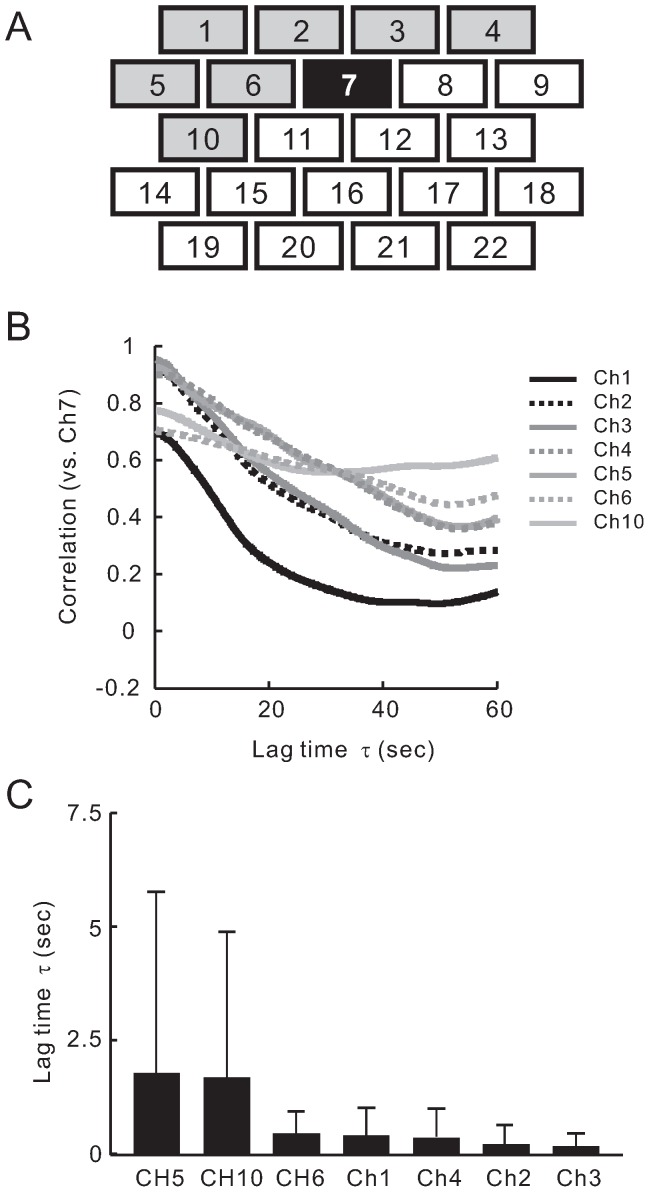
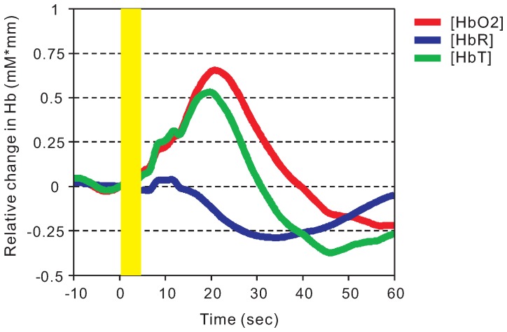
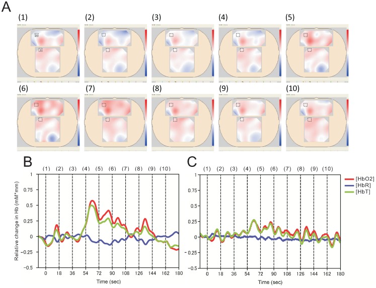
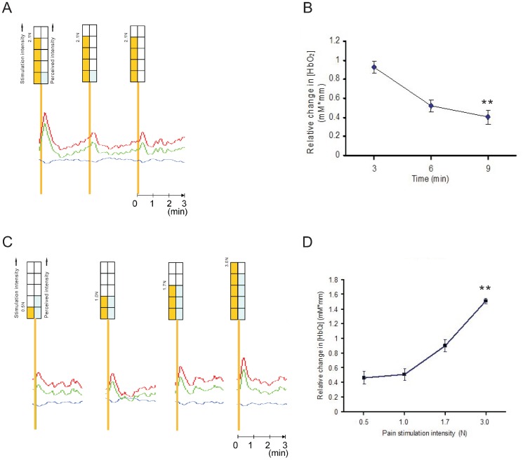
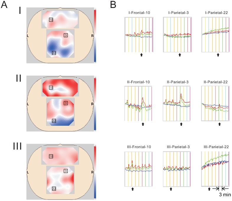
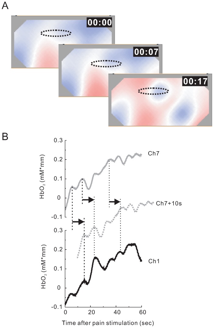
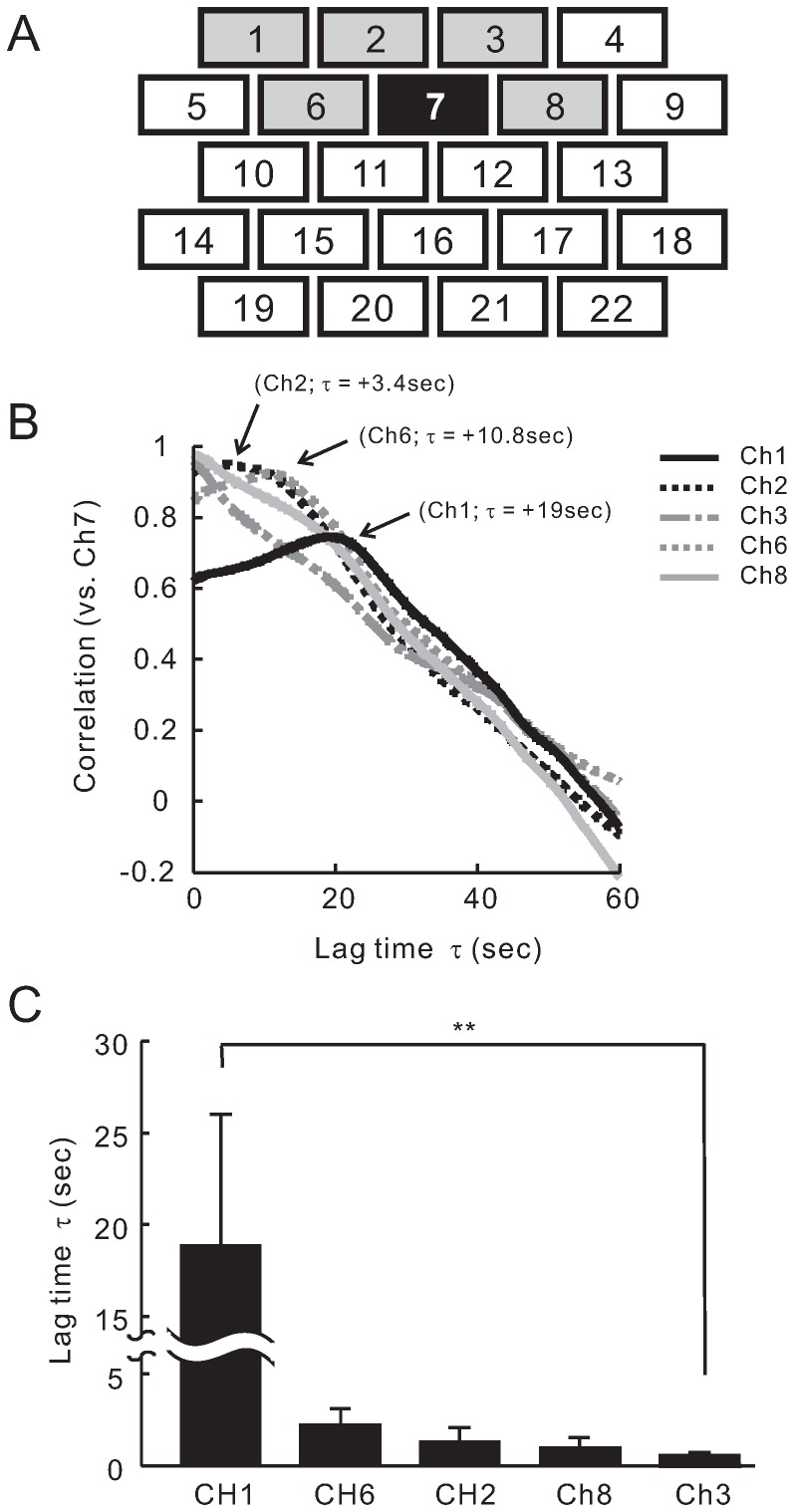
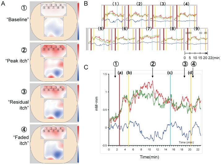
Pain and itch are closely related sensations, yet qualitatively quite distinct. Despite recent advances in brain imaging techniques, identifying the differences between pain and itch signals in the brain cortex is difficult due to continuous temporal and spatial changes in the signals. The high spatial resolution of positron emission tomography (PET) and functional magnetic resonance imaging (fMRI) has substantially advanced research of pain and itch, but these are uncomfortable because of expensiveness, importability and the limited operation in the shielded room. Here, we used near infrared spectroscopy (NIRS), which has more conventional usability. NIRS can be used to visualize dynamic changes in oxygenated hemoglobin and deoxyhemoglobin concentrations in the capillary networks near activated neural circuits in real-time as well as fMRI. We observed distinct activation patterns in the frontal cortex for acute pain and histamine-induced itch. The prefrontal cortex exhibited a pain-related and itch-related activation pattern of blood flow in each subject. Although it looked as though that activation pattern for pain and itching was different in each subject, further cross correlation analysis of NIRS signals between each channels showed an overall agreement with regard to prefrontal area involvement. As a result, pain-related and itch-related blood flow responses (delayed responses in prefrontal area) were found to be clearly different between pain (τ = +18.7 sec) and itch (τ = +0.63 sec) stimulation. This is the first pilot study to demonstrate the temporal and spatial separation of a pain-induced blood flow and an itch-induced blood flow in human cortex during information processing.
疼痛和瘙痒是密切相关的感觉,但在性质上却截然不同。尽管脑成像技术最近取得了进展,但由于信号在时间和空间上的持续变化,识别大脑皮层中疼痛和瘙痒信号之间的差异仍然很困难。正电子发射断层扫描(PET)和功能磁共振成像(fMRI)的高空间分辨率极大地推动了对疼痛和瘙痒的研究,但由于费用高昂、难以引入以及在屏蔽室中的操作受限,这些方法并不舒适。在这里,我们使用了更具通用性的近红外光谱(NIRS)。NIRS可以像fMRI一样实时可视化激活神经回路附近毛细血管网络中氧合血红蛋白和脱氧血红蛋白浓度的动态变化。我们观察到急性疼痛和组胺诱导的瘙痒在额叶皮层有明显的激活模式。前额叶皮层在每个受试者中都表现出与疼痛相关和与瘙痒相关的血流激活模式。尽管每个受试者的疼痛和瘙痒激活模式看起来不同,但对每个通道之间的NIRS信号进行进一步的互相关分析表明,在前额叶区域的参与方面总体上是一致的。结果发现,在疼痛(τ = +18.7秒)和瘙痒(τ = +0.63秒)刺激期间,与疼痛相关和与瘙痒相关的血流反应(前额叶区域的延迟反应)明显不同。这是第一项初步研究,证明了在信息处理过程中,人类皮层中疼痛诱导的血流和瘙痒诱导的血流在时间和空间上的分离。