Frick Andreas, Gingnell Malin, Marquand Andre F, Howner Katarina, Fischer Håkan, Kristiansson Marianne, Williams Steven C R, Fredrikson Mats, Furmark Tomas
Department of Psychology, Uppsala University, Uppsala, Sweden.
Department of Psychology, Uppsala University, Uppsala, Sweden; Department of Women's and Children's Health, Uppsala University, Uppsala, Sweden.
Behav Brain Res. 2014 Feb 1;259:330-5. doi: 10.1016/j.bbr.2013.11.003. Epub 2013 Nov 13.
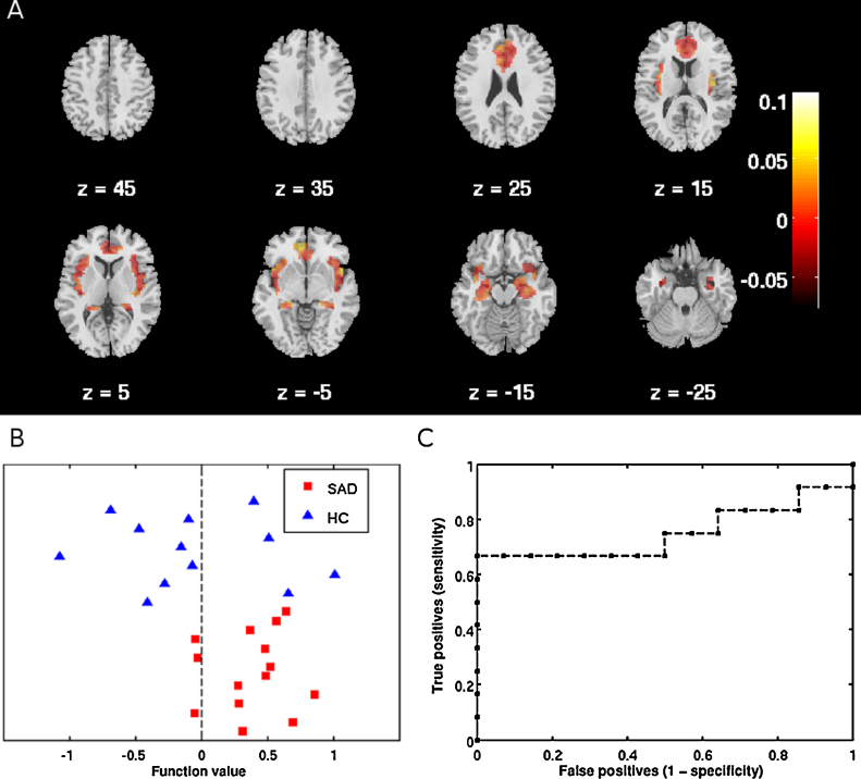
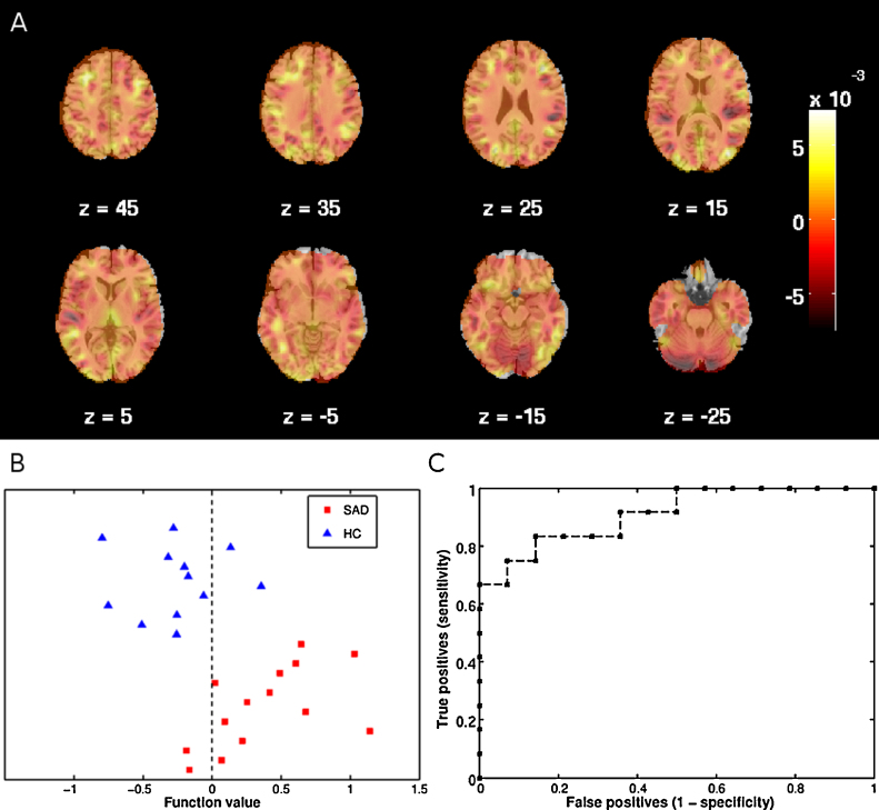
Functional neuroimaging of social anxiety disorder (SAD) support altered neural activation to threat-provoking stimuli focally in the fear network, while structural differences are distributed over the temporal and frontal cortices as well as limbic structures. Previous neuroimaging studies have investigated the brain at the voxel level using mass-univariate methods which do not enable detection of more complex patterns of activity and structural alterations that may separate SAD from healthy individuals. Support vector machine (SVM) is a supervised machine learning method that capitalizes on brain activation and structural patterns to classify individuals. The aim of this study was to investigate if it is possible to discriminate SAD patients (n=14) from healthy controls (n=12) using SVM based on (1) functional magnetic resonance imaging during fearful face processing and (2) regional gray matter volume. Whole brain and region of interest (fear network) SVM analyses were performed for both modalities. For functional scans, significant classifications were obtained both at whole brain level and when restricting the analysis to the fear network while gray matter SVM analyses correctly classified participants only when using the whole brain search volume. These results support that SAD is characterized by aberrant neural activation to affective stimuli in the fear network, while disorder-related alterations in regional gray matter volume are more diffusely distributed over the whole brain. SVM may thus be useful for identifying imaging biomarkers of SAD.
社交焦虑障碍(SAD)的功能神经影像学研究表明,在恐惧网络中,对引发威胁的刺激,其神经激活发生了改变,而结构差异则分布在颞叶和额叶皮质以及边缘结构。以往的神经影像学研究使用单变量方法在体素水平上研究大脑,这种方法无法检测到可能将社交焦虑障碍患者与健康个体区分开来的更复杂的活动和结构改变模式。支持向量机(SVM)是一种监督式机器学习方法,它利用大脑激活和结构模式对个体进行分类。本研究的目的是调查是否有可能基于(1)恐惧面孔加工过程中的功能磁共振成像和(2)局部灰质体积,使用支持向量机将社交焦虑障碍患者(n = 14)与健康对照组(n = 12)区分开来。对这两种模式都进行了全脑和感兴趣区域(恐惧网络)的支持向量机分析。对于功能扫描,在全脑水平以及将分析限制在恐惧网络时都获得了显著的分类结果,而灰质支持向量机分析仅在使用全脑搜索体积时才正确分类了参与者。这些结果支持社交焦虑障碍的特征是恐惧网络中对情感刺激的异常神经激活,而区域灰质体积的障碍相关改变在全脑中分布更广泛。因此,支持向量机可能有助于识别社交焦虑障碍的影像学生物标志物。