Department of Clinical Sciences Malmö, Molecular Memory Research Unit, Lund University, The Wallenberg Laboratory 2nd floor, Inga Marie Nilssons gata, entrance 53, Skåne University Hospital, Malmö, 205 02, Sweden.
Acta Neuropathol Commun. 2013 May 9;1(1):7. doi: 10.1186/2051-5960-1-7.
Neuron Glial 2 (NG2) cells are glial cells known to serve as oligodendrocyte progenitors as well as modulators of the neuronal network. Altered NG2 cell morphology and up-regulation as well as increased shedding of the proteoglycan NG2 expressed on the cell surface have been described in rodent models of brain injury. Here we describe alterations in the human NG2 cell population in response to pathological changes characteristic of Alzheimer's disease (AD).
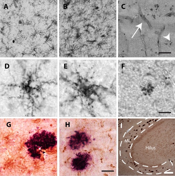
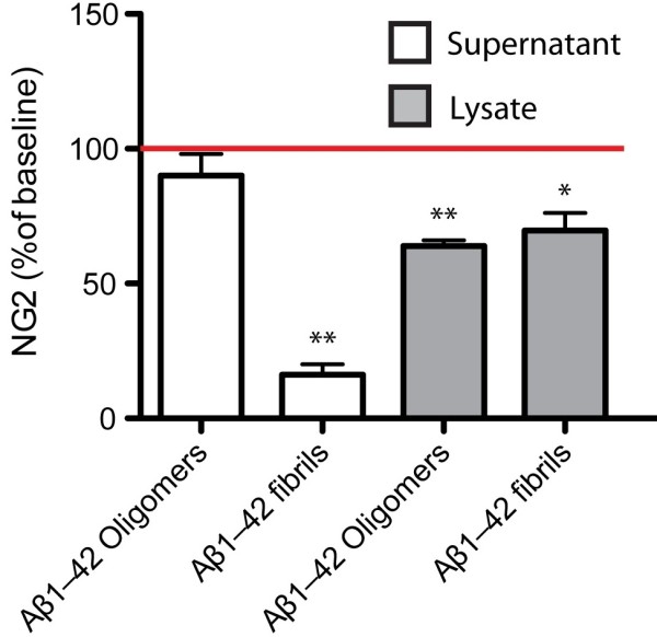
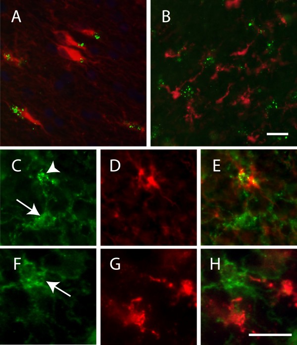
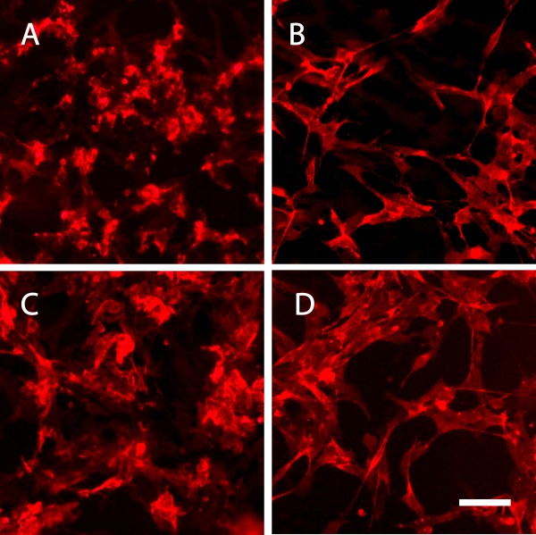
Immunohistological stainings of postmortem brain specimens from clinically diagnosed and postmortem verified AD patients and non-demented controls revealed reduced NG2 immunoreactivity as well as large numbers of NG2 positive astrocytes in individuals with high amyloid beta plaque load. Since fibrillar amyloid beta (Aβ)1-42 is the major component of AD-related senile plaques, we exposed human NG2 cells to oligomer- and fibril enriched preparations of Aβ1-42. We found that both oligomeric and fibrillar Aβ1-42 induced changes in NG2 cell morphology. Further, in vitro exposure to fibrillar Aβ1-42 decreased the NG2 concentrations in both cell lysates and supernatants. Interestingly, we also found significantly decreased levels of soluble NG2 in the cerebrospinal fluid (CSF) from clinically diagnosed AD patients compared to non-demented individuals. Additionally, the CSF NG2 levels were found to significantly correlate with the core AD biomarkers Aß1-42, T-tau and P-tau.
Our results demonstrate major alterations in the NG2 cell population in relation to AD pathology which highlights the NG2 cell population as a new attractive research target in the search for cellular mechanisms associated with AD pathogenesis.
神经胶质细胞 2(NG2)细胞是一种神经胶质细胞,已知其作为少突胶质细胞前体以及神经元网络的调节剂。在啮齿动物脑损伤模型中,已经描述了 NG2 细胞形态的改变以及表面表达的蛋白聚糖 NG2 的上调和脱落增加。在这里,我们描述了人类 NG2 细胞群体对阿尔茨海默病(AD)特征性病理变化的反应的改变。
对临床诊断和死后证实的 AD 患者和非痴呆对照的死后脑标本进行免疫组织化学染色显示,NG2 免疫反应性降低,并且在高淀粉样β斑块负荷的个体中存在大量 NG2 阳性星形胶质细胞。由于纤维状淀粉样β(Aβ)1-42 是 AD 相关老年斑的主要成分,我们将人类 NG2 细胞暴露于寡聚体和富含纤维的 Aβ1-42 制剂中。我们发现,寡聚体和纤维状 Aβ1-42 均可诱导 NG2 细胞形态发生变化。此外,体外暴露于纤维状 Aβ1-42 可降低细胞裂解物和上清液中 NG2 的浓度。有趣的是,我们还发现与非痴呆个体相比,来自临床诊断为 AD 患者的脑脊液(CSF)中可溶性 NG2 的水平显著降低。此外,CSF NG2 水平与 AD 的核心生物标志物 Aß1-42、T-tau 和 P-tau 呈显著相关性。
我们的结果表明,NG2 细胞群体在 AD 病理学方面发生了重大改变,这突显了 NG2 细胞群体作为与 AD 发病机制相关的细胞机制的新的有吸引力的研究目标。