Arthritis Res Ther. 2013 Sep 20;15(5):R129. doi: 10.1186/ar4309.
Treatment of chondral injuries remains a major issue despite the many advances made in cartilage repair techniques. Although it has been postulated that the use of marrow stimulation in combination with cell-based therapy may provide superior outcome, this has yet to be demonstrated. A pilot study was thus conducted to determine if bone marrow derived mesenchymal stromal cells (BM-MSCs) have modulatory effects on the repair outcomes of bone marrow stimulation (BMS) techniques.
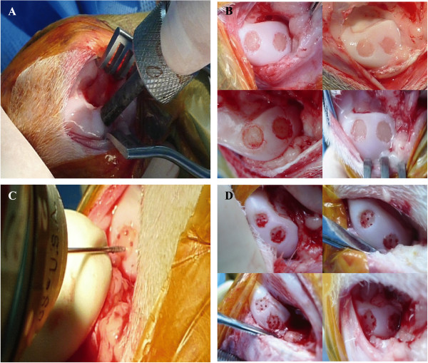
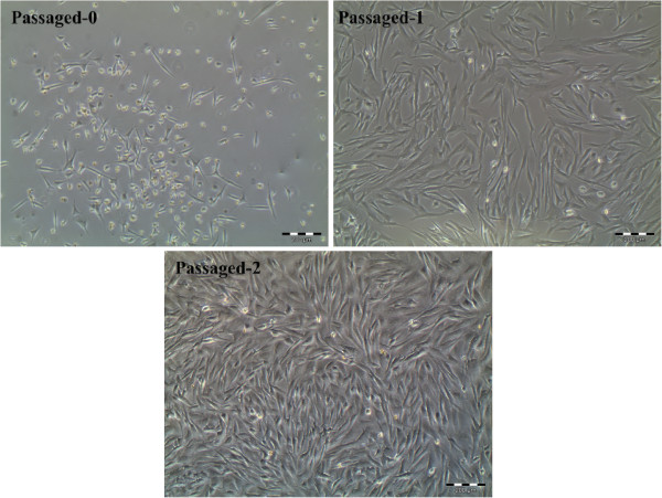
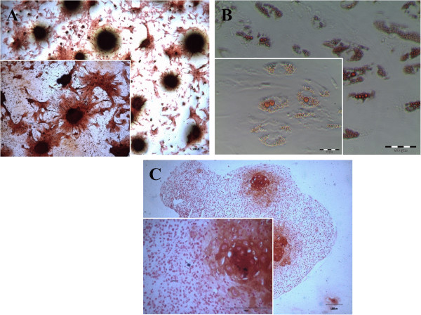
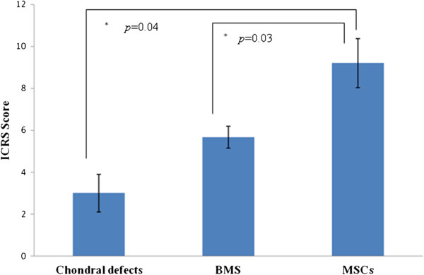
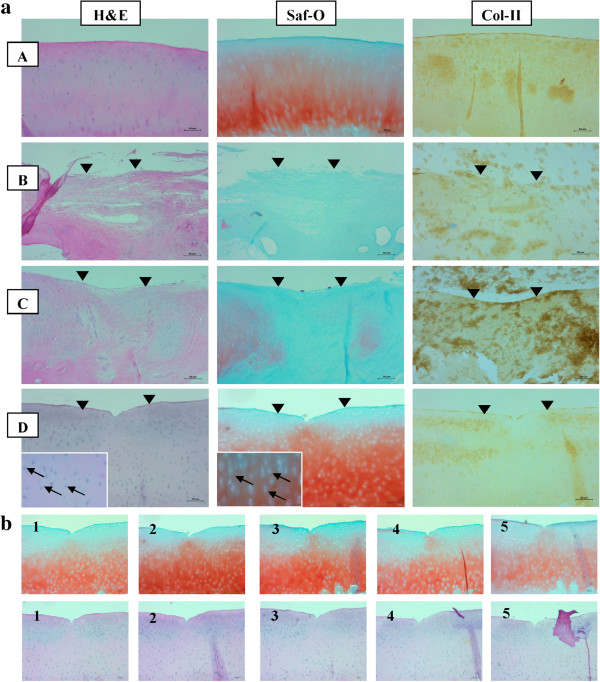
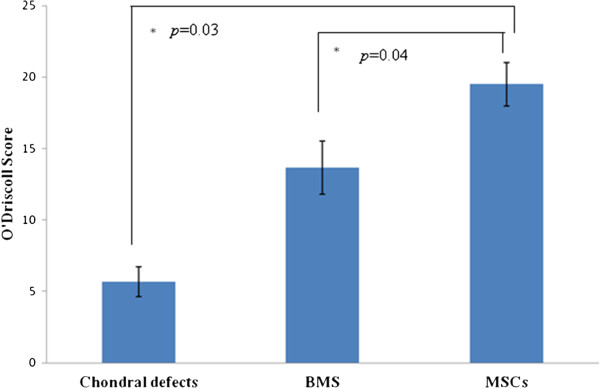
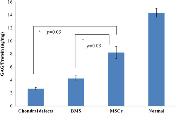
Two full-thickness chondral 5 mm diameter defects were created in tandem on the medial condyle of left stifle joints of 18 Boer caprine (N = 18). Goats were then divided equally into three groups. Simultaneously, bone marrow aspirates were taken from the iliac crests from the goats in Group 1 and were sent for BM-MSC isolation and expansion in vitro. Six weeks later, BMS surgery, which involves subchondral drilling at the defect sites, was performed. After two weeks, the knees in Group 1 were given autologous intra-articular BM-MSCs (N = 6). In Group 2, although BMS was performed there were no supplementations provided. In Group 3, no intervention was administered. The caprines were sacrificed after six months. Repairs were evaluated using macroscopic assessment through the International Cartilage Repair Society (ICRS) scoring, histologic grading by O'Driscoll score, biochemical assays for glycosaminoglycans (GAGs) and gene expressions for aggrecan, collagen II and Sox9.
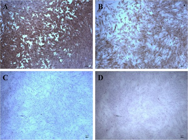
Histological and immunohistochemical analyses demonstrated hyaline-like cartilage regeneration in the transplanted sites particularly in Group 1. In contrast, tissues in Groups 2 and 3 demonstrated mainly fibrocartilage. The highest ICRS and O'Driscoll scorings was also observed in Group 1, while the lowest score was seen in Group 3. Similarly, the total GAG/total protein as well as chondrogenic gene levels were expressed in the same order, that is highest in Group 1 while the lowest in Group three. Significant differences between these 3 groups were observed (P <0.05).
This study suggests that supplementing intra-articular injections of BM-MSCs following BMS knee surgery provides superior cartilage repair outcomes.
尽管在软骨修复技术方面取得了许多进展,但软骨损伤的治疗仍然是一个主要问题。虽然有人推测,骨髓刺激与细胞治疗相结合可能会提供更好的结果,但这尚未得到证实。因此,进行了一项初步研究,以确定骨髓来源的间充质基质细胞(BM-MSCs)是否对骨髓刺激(BMS)技术的修复结果有调节作用。
在 18 只布尔山羊的左膝关节内侧髁上同时创建了两个 5 毫米直径的全层软骨缺损(N=18)。然后将山羊平均分为三组。同时,从组 1 中的山羊的髂嵴采集骨髓抽吸物,并送往体外进行 BM-MSC 分离和扩增。六周后,在缺陷部位进行 BMS 手术,包括软骨下钻孔。两周后,组 1 的膝关节给予自体关节内 BM-MSCs(N=6)。在组 2 中,虽然进行了 BMS 但没有提供补充。在组 3 中,未进行干预。六个月后处死山羊。使用国际软骨修复协会(ICRS)评分进行宏观评估、O'Driscoll 评分进行组织学分级、糖胺聚糖(GAG)生化测定和聚集蛋白聚糖、胶原 II 和 Sox9 的基因表达来评估修复情况。
组织学和免疫组织化学分析表明,移植部位有透明软骨样软骨再生,特别是在组 1 中。相比之下,组 2 和组 3 的组织主要为纤维软骨。组 1 的 ICRS 和 O'Driscoll 评分最高,而组 3 的评分最低。同样,总 GAG/总蛋白以及软骨形成基因水平也以相同的顺序表达,即组 1 最高,组 3 最低。这三组之间观察到显著差异(P<0.05)。
本研究表明,BMS 膝关节手术后关节内注射 BM-MSCs 可提供更好的软骨修复效果。