Fan Jui-Lin, Bourdillon Nicolas, Kayser Bengt
Institute of Sports Sciences, Faculty of Biology and Medicine, University of Lausanne Lausanne, Switzerland ; Lemanic Doctoral School of Neuroscience, University of Lausanne Lausanne, Switzerland.
Physiol Rep. 2013 Aug;1(3):e00066. doi: 10.1002/phy2.66. Epub 2013 Aug 28.
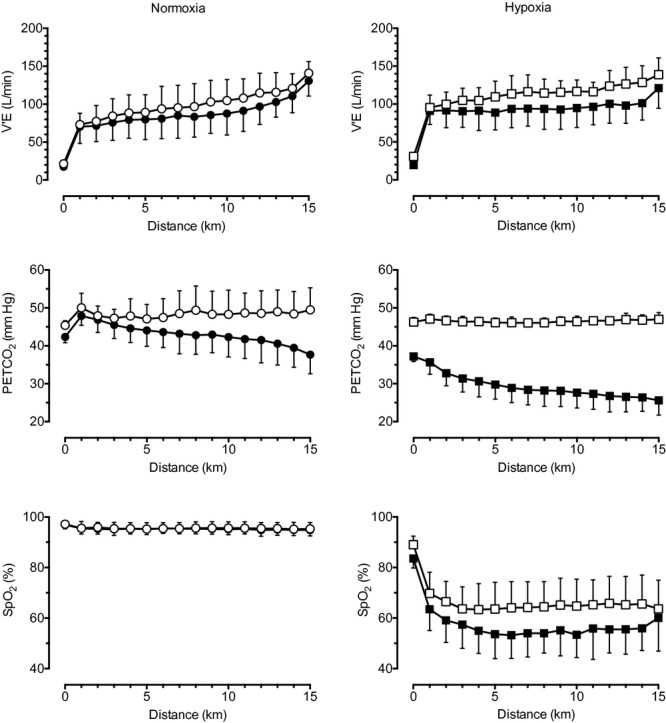
During heavy exercise, hyperventilation-induced hypocapnia leads to cerebral vasoconstriction, resulting in a reduction in cerebral blood flow (CBF). A reduction in CBF would impair cerebral O2 delivery and potentially account for reduced exercise performance in hypoxia. We tested the hypothesis that end-tidal Pco2 (PETCO2) clamping in hypoxic exercise would prevent the hypocapnia-induced reduction in CBF during heavy exercise, thus improving exercise performance. We measured PETCO2, middle cerebral artery velocity (MCAv; index of CBF), prefrontal cerebral cortex oxygenation (cerebral O2Hb; index of cerebral oxygenation), cerebral O2 delivery (DO2), and leg muscle oxygenation (muscle O2Hb) in 10 healthy men (age 27 ± 7 years; VO2max 63.3 ± 6.6 mL/kg/min; mean ± SD) during simulated 15-km time trial cycling (TT) in normoxia and hypoxia (FIO2 = 0.10) with and without CO2 clamping. During exercise, hypoxia elevated MCAv and lowered cerebral O2Hb, cerebral DO2, and muscle O2Hb (P < 0.001). CO2 clamping elevated PETCO2 and MCAv during exercise in both normoxic and hypoxic conditions (P < 0.001 and P = 0.024), but had no effect on either cerebral and muscle O2Hb (P = 0.118 and P = 0.124). Nevertheless, CO2 clamping elevated cerebral DO2 during TT in both normoxic and hypoxic conditions (P < 0.001). CO2 clamping restored cerebral DO2 to normoxic values during TT in hypoxia and tended to have a greater effect on TT performance in hypoxia compared to normoxia (P = 0.097). However, post hoc analysis revealed no effect of CO2 clamping on TT performance either in normoxia (P = 0.588) or in hypoxia (P = 0.108). Our findings confirm that the hyperventilation-induced hypocapnia and the subsequent drop in cerebral oxygenation are unlikely to be the cause of the reduced endurance exercise performance in hypoxia.
在剧烈运动期间,过度通气引起的低碳酸血症会导致脑血管收缩,从而使脑血流量(CBF)减少。脑血流量减少会损害脑氧输送,并可能导致低氧环境下运动表现下降。我们检验了这样一个假设:在低氧运动中进行呼气末二氧化碳分压(PETCO2)钳夹可防止剧烈运动期间低碳酸血症引起的脑血流量减少,从而改善运动表现。我们测量了10名健康男性(年龄27±7岁;最大摄氧量63.3±6.6 mL/kg/min;平均值±标准差)在常氧和低氧(吸入氧分数FIO2 = 0.10)状态下进行模拟15公里计时赛骑行(TT)时的PETCO2、大脑中动脉血流速度(MCAv;脑血流量指标)、前额叶皮层氧合(脑氧合血红蛋白;脑氧合指标)、脑氧输送(DO2)和腿部肌肉氧合(肌肉氧合血红蛋白),实验分为有和没有二氧化碳钳夹两种情况。运动期间,低氧状态会使MCAv升高,并降低脑氧合血红蛋白、脑DO2和肌肉氧合血红蛋白(P < 0.001)。在常氧和低氧条件下,运动期间二氧化碳钳夹都会使PETCO2和MCAv升高(P < 0.001和P = 0.02),但对脑和肌肉氧合血红蛋白均无影响(P = 0.118和P = 0.124)。尽管如此,在常氧和低氧条件下,TT期间二氧化碳钳夹都会使脑DO2升高(P < 0.001)。在低氧状态下的TT期间,二氧化碳钳夹可使脑DO恢复到常氧值,并且与常氧相比,对低氧状态下的TT表现影响更大(P = 0.097)。然而,事后分析显示,二氧化碳钳夹在常氧(P = 0.588)或低氧(P = 0.108)状态下对TT表现均无影响。我们的研究结果证实,过度通气引起的低碳酸血症以及随后的脑氧合下降不太可能是低氧环境下耐力运动表现下降的原因。