Department of Physiology and Biophysics and Center for Cardiovascular Research, College of Medicine, University of Illinois at Chicago Chicago, IL USA.
Front Physiol. 2013 Nov 20;4:336. doi: 10.3389/fphys.2013.00336. eCollection 2013.
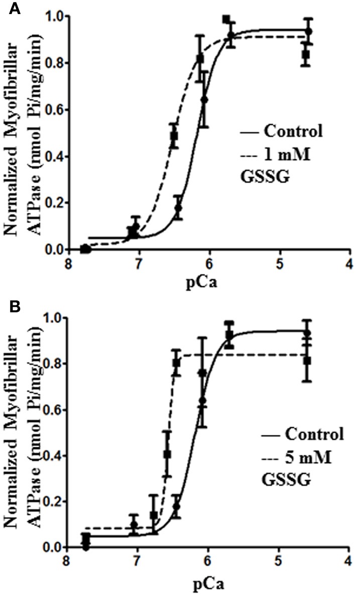
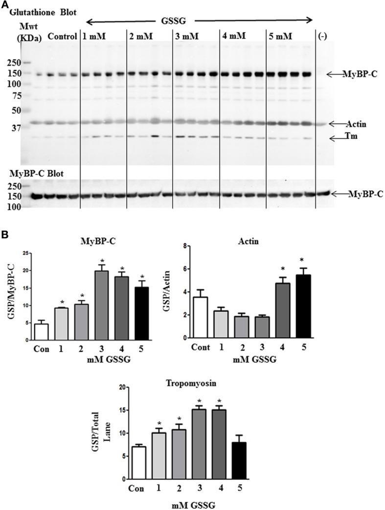
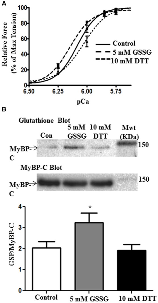
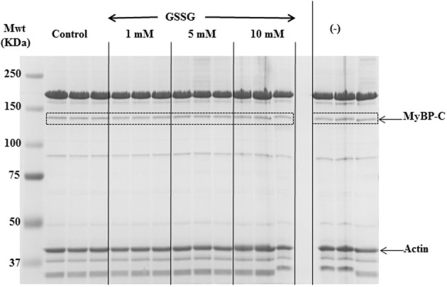
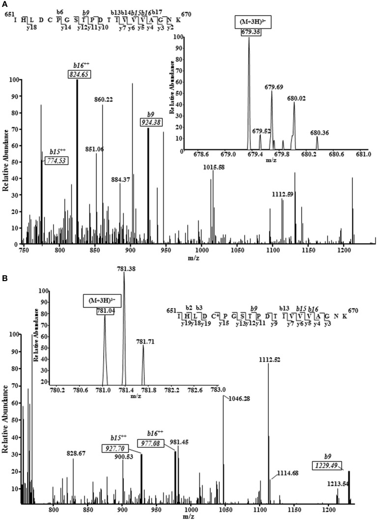
Our previous studies demonstrated a relation between glutathionylation of cardiac myosin binding protein C (cMyBP-C) and diastolic dysfunction in a hypertensive mouse model stressed by treatment with salt, deoxycorticosterone acetate, and unilateral nephrectomy. Although these results strongly indicated an important role for S-glutathionylation of myosin binding protein C as a modifier of myofilament function, indirect effects of other post-translational modifications may have occurred. Moreover, we did not determine the sites of thiol modification by glutathionylation. To address these issues, we developed an in vitro method to mimic the in situ S-glutathionylation of myofilament proteins and determined direct functional effects and sites of oxidative modification employing Western blotting and mass spectrometry. We induced glutathionylation in vitro by treatment of isolated myofibrils and detergent extracted fiber bundles (skinned fibers) with oxidized glutathione (GSSG). Immuno-blotting results revealed increased glutathionylation with GSSG treatment of a protein band around 140 kDa. Using tandem mass spectrometry, we identified the 140 kDa band as cMyBP-C and determined the sites of glutathionylation to be at cysteines 655, 479, and 627. Determination of the relation between Ca(2+)-activation of myofibrillar acto-myosin ATPase rate demonstrated an increased Ca(2+)-sensitivity induced by the S-glutathionylation. Force generating skinned fiber bundles also showed an increase in Ca-sensitivity when treated with oxidized glutathione, which was reversed with the reducing agent, dithiothreitol (DTT). Our data demonstrate that a specific and direct effect of S-glutathionylation of myosin binding protein C is a significant increase in myofilament Ca(2+)-sensitivity. Our data also provide new insights into the functional significance of oxidative modification of myosin binding protein C and the potential role of domains not previously considered to be functionally significant as controllers of myofilament Ca(2+)-responsiveness and dynamics.
我们之前的研究表明,在盐皮质酮、脱氧皮质酮和单侧肾切除处理的高血压小鼠模型中,肌球蛋白结合蛋白 C(cMyBP-C)的谷胱甘肽化与舒张功能障碍之间存在关系。尽管这些结果强烈表明肌球蛋白结合蛋白 C 的 S-谷胱甘肽化作为肌丝功能调节剂的重要作用,但可能发生了其他翻译后修饰的间接影响。此外,我们没有确定通过谷胱甘肽化修饰的硫醇修饰位点。为了解决这些问题,我们开发了一种体外方法来模拟肌球蛋白蛋白丝蛋白的原位 S-谷胱甘肽化,并通过 Western blot 和质谱法确定直接功能影响和氧化修饰的位点。我们通过用氧化型谷胱甘肽(GSSG)处理分离的肌原纤维和去污剂提取的纤维束(去细胞纤维)在体外诱导谷胱甘肽化。免疫印迹结果显示,用 GSSG 处理后,约 140 kDa 的蛋白质带的谷胱甘肽化增加。通过串联质谱法,我们确定 140 kDa 带为 cMyBP-C,并确定谷胱甘肽化的位点为半胱氨酸 655、479 和 627。肌球蛋白丝肌球蛋白 ATP 酶活性的 Ca2+激活关系的测定表明,S-谷胱甘肽化诱导 Ca2+敏感性增加。用氧化型谷胱甘肽处理产生力的去细胞纤维束也显示 Ca 敏感性增加,用还原剂二硫苏糖醇(DTT)处理可逆转。我们的数据表明,肌球蛋白结合蛋白 C 的 S-谷胱甘肽化的一个特定和直接作用是肌丝 Ca2+敏感性的显著增加。我们的数据还为肌球蛋白结合蛋白 C 的氧化修饰的功能意义以及以前认为在控制肌丝 Ca2+反应性和动力学方面没有功能意义的区域的潜在作用提供了新的见解。