• 文献检索
  • 文档翻译
  • 深度研究
  • 学术资讯
  • Suppr Zotero 插件Zotero 插件
  • 邀请有礼
  • 套餐&价格
  • 历史记录
应用&插件
Suppr Zotero 插件Zotero 插件浏览器插件Mac 客户端Windows 客户端微信小程序
定价
高级版会员购买积分包购买API积分包
服务
文献检索文档翻译深度研究API 文档MCP 服务
关于我们
关于 Suppr公司介绍联系我们用户协议隐私条款
关注我们

Suppr 超能文献

核心技术专利:CN118964589B侵权必究
粤ICP备2023148730 号-1Suppr @ 2026

文献检索

告别复杂PubMed语法,用中文像聊天一样搜索,搜遍4000万医学文献。AI智能推荐,让科研检索更轻松。

立即免费搜索

文件翻译

保留排版,准确专业,支持PDF/Word/PPT等文件格式,支持 12+语言互译。

免费翻译文档

深度研究

AI帮你快速写综述,25分钟生成高质量综述,智能提取关键信息,辅助科研写作。

立即免费体验

RNA 结合蛋白 RBPMS 是哺乳动物视网膜神经节细胞的选择性标志物。

The RNA binding protein RBPMS is a selective marker of ganglion cells in the mammalian retina.

机构信息

Department of Neurobiology, David Geffen School of Medicine at Los Angeles, University of California at Los Angeles, Los Angeles, California, 90095-1763.

出版信息

J Comp Neurol. 2014 Apr 15;522(6):1411-43. doi: 10.1002/cne.23521.

DOI:10.1002/cne.23521
PMID:24318667
原文链接:https://pmc.ncbi.nlm.nih.gov/articles/PMC3959221/
Abstract

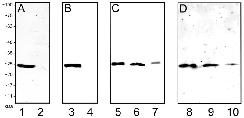
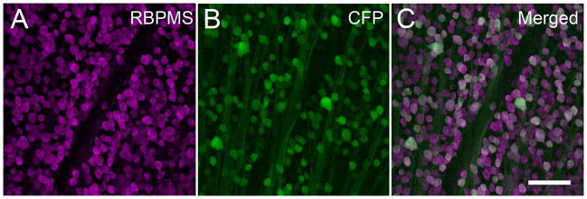
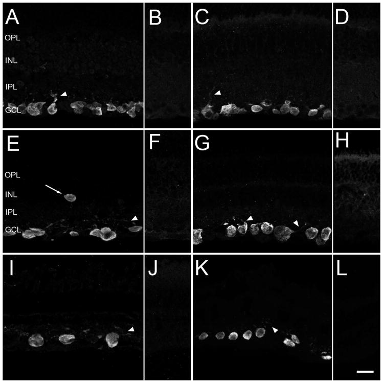
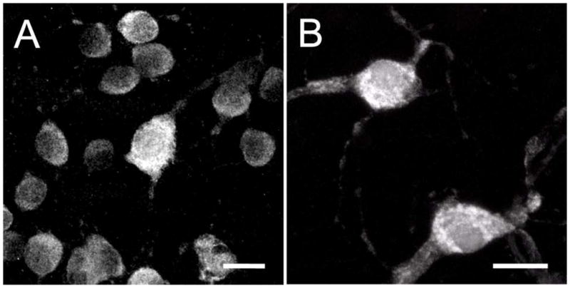
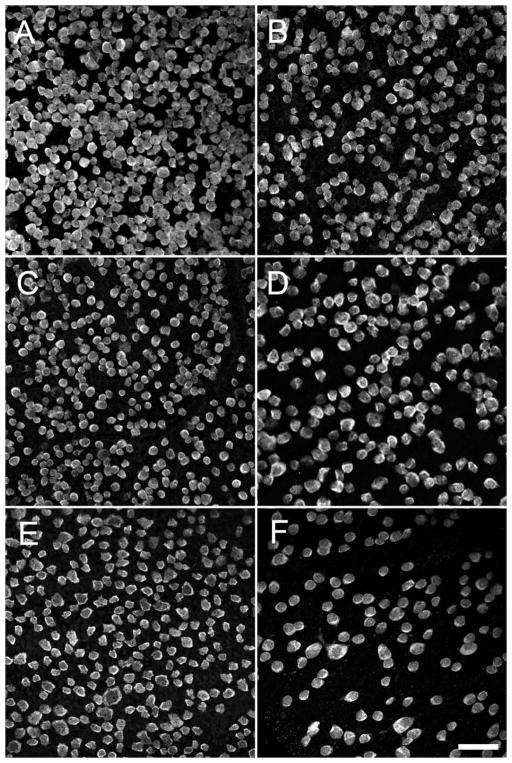
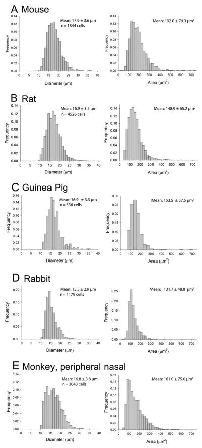
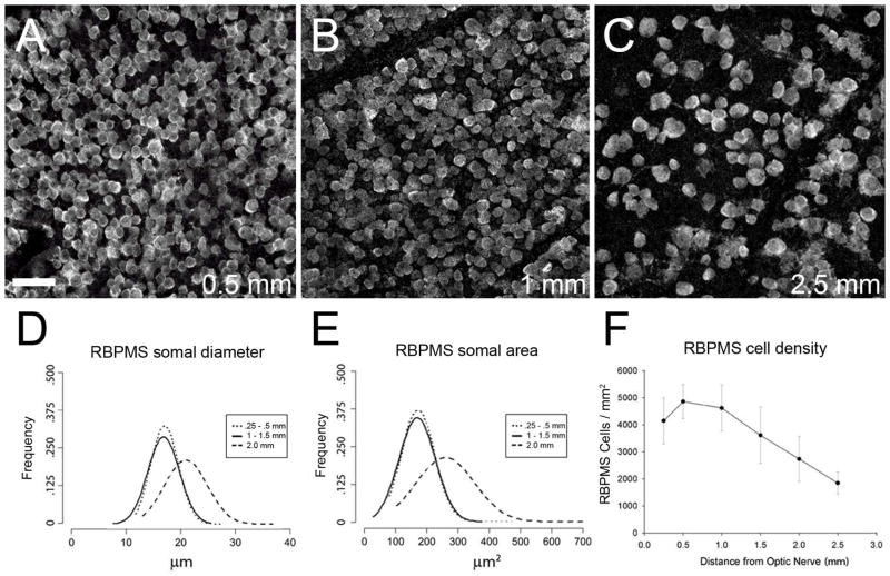
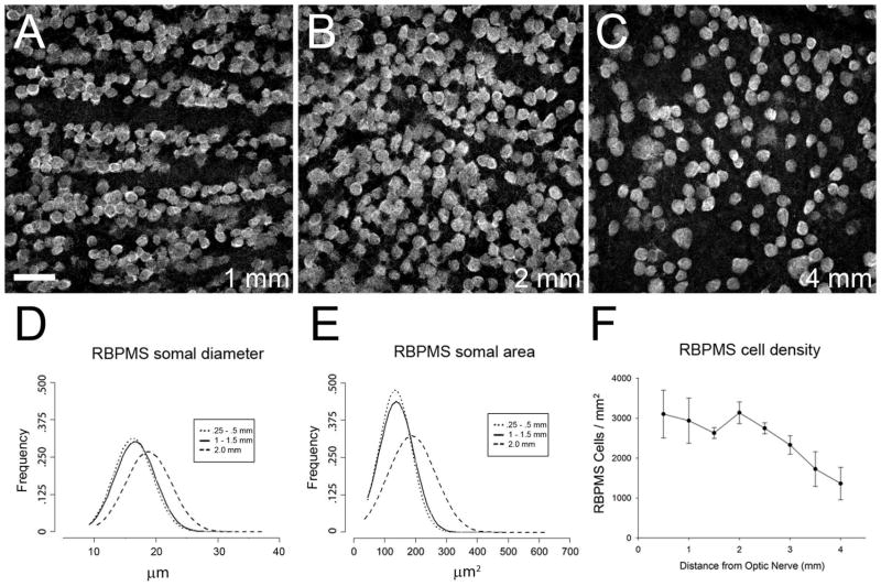
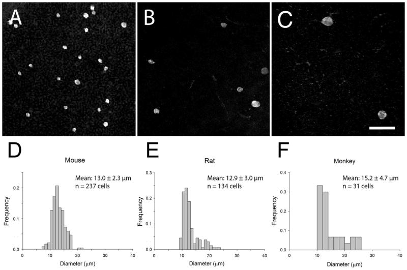
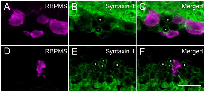
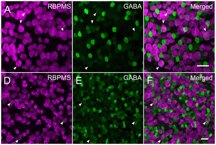
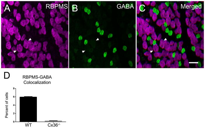
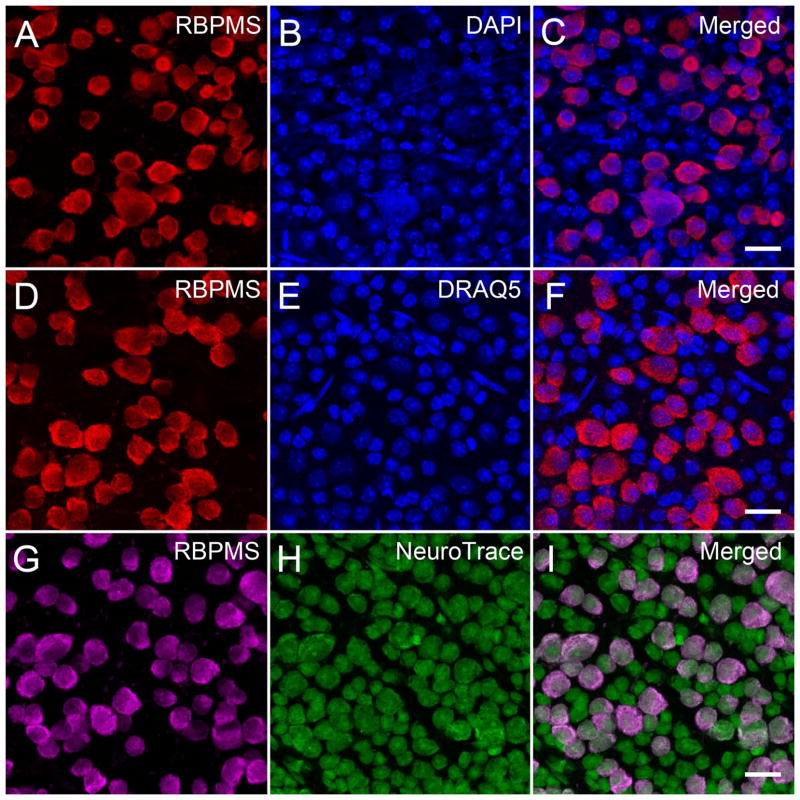
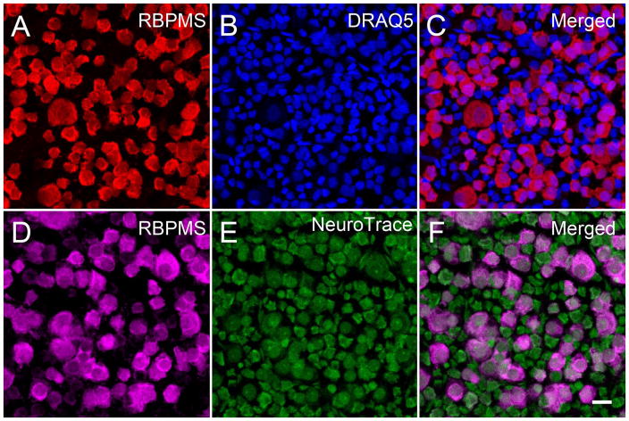
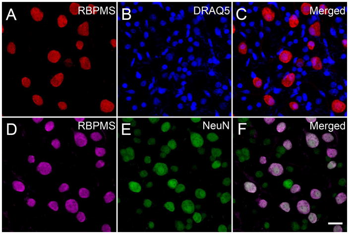
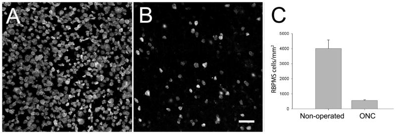
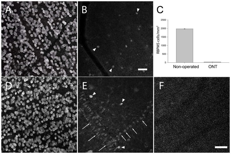
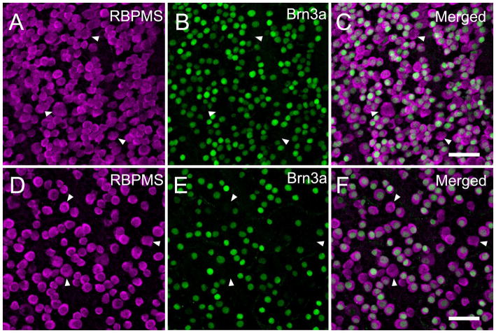
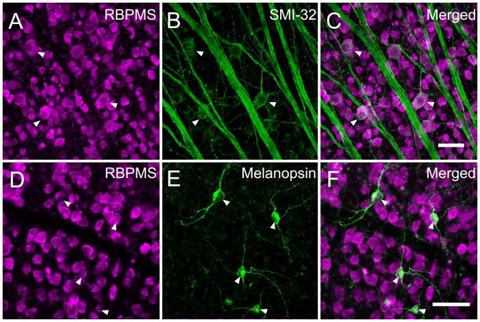
There are few neurochemical markers that reliably identify retinal ganglion cells (RGCs), which are a heterogeneous population of cells that integrate and transmit the visual signal from the retina to the central visual nuclei. We have developed and characterized a new set of affinity-purified guinea pig and rabbit antibodies against RNA-binding protein with multiple splicing (RBPMS). On western blots these antibodies recognize a single band at 〜24 kDa, corresponding to RBPMS, and they strongly label RGC and displaced RGC (dRGC) somata in mouse, rat, guinea pig, rabbit, and monkey retina. RBPMS-immunoreactive cells and RGCs identified by other techniques have a similar range of somal diameters and areas. The density of RBPMS cells in mouse and rat retina is comparable to earlier semiquantitative estimates of RGCs. RBPMS is mainly expressed in medium and large DAPI-, DRAQ5-, NeuroTrace- and NeuN-stained cells in the ganglion cell layer (GCL), and RBPMS is not expressed in syntaxin (HPC-1)-immunoreactive cells in the inner nuclear layer (INL) and GCL, consistent with their identity as RGCs, and not displaced amacrine cells. In mouse and rat retina, most RBPMS cells are lost following optic nerve crush or transection at 3 weeks, and all Brn3a-, SMI-32-, and melanopsin-immunoreactive RGCs also express RBPMS immunoreactivity. RBPMS immunoreactivity is localized to cyan fluorescent protein (CFP)-fluorescent RGCs in the B6.Cg-Tg(Thy1-CFP)23Jrs/J mouse line. These findings show that antibodies against RBPMS are robust reagents that exclusively identify RGCs and dRGCs in multiple mammalian species, and they will be especially useful for quantification of RGCs.

摘要

几乎没有神经化学标记物能够可靠地识别视网膜神经节细胞(RGC),RGC 是一个异质细胞群体,它们整合并将视觉信号从视网膜传递到中央视觉核。我们开发并表征了一组新的针对 RNA 结合蛋白多剪接(RBPMS)的亲和纯化豚鼠和兔抗体。在 Western blot 上,这些抗体识别出一个约 24 kDa 的单一条带,对应于 RBPMS,并且它们强烈标记小鼠、大鼠、豚鼠、兔和猴视网膜中的 RGC 和移位的 RGC(dRGC)体。通过其他技术鉴定的 RBPMS 免疫反应性细胞和 RGC 具有相似的体直径和面积范围。在小鼠和大鼠视网膜中,RBPMS 细胞的密度与以前对 RGC 的半定量估计相当。RBPMS 主要在神经追踪剂(NeuroTrace)、DAPI、DRAQ5 和 NeuN 染色的中等和大直径 DAPI、DRAQ5、NeuroTrace 和 NeuN 染色的细胞层(GCL)中表达,并且 RBPMS 不在内核层(INL)和 GCL 中的 syntaxin(HPC-1)免疫反应性细胞中表达,与它们作为 RGC 的身份一致,而不是移位的无长突细胞。在小鼠和大鼠视网膜中,视神经挤压或 3 周时横断后,大多数 RBPMS 细胞丢失,所有 Brn3a、SMI-32 和黑视蛋白免疫反应性 RGC 也表达 RBPMS 免疫反应性。RBPMS 免疫反应性定位于 B6.Cg-Tg(Thy1-CFP)23Jrs/J 小鼠系中 CFP 荧光 RGC 的青色荧光蛋白(CFP)荧光。这些发现表明,针对 RBPMS 的抗体是一种强大的试剂,可特异性识别多种哺乳动物物种中的 RGC 和 dRGC,它们对于 RGC 的定量特别有用。

https://cdn.ncbi.nlm.nih.gov/pmc/blobs/78fc/3959221/790258098236/nihms548668f19.jpg
https://cdn.ncbi.nlm.nih.gov/pmc/blobs/78fc/3959221/148a3377e9f0/nihms548668f1.jpg
https://cdn.ncbi.nlm.nih.gov/pmc/blobs/78fc/3959221/8423841f3d81/nihms548668f2.jpg
https://cdn.ncbi.nlm.nih.gov/pmc/blobs/78fc/3959221/9c428f969501/nihms548668f3.jpg
https://cdn.ncbi.nlm.nih.gov/pmc/blobs/78fc/3959221/24c071985f29/nihms548668f4.jpg
https://cdn.ncbi.nlm.nih.gov/pmc/blobs/78fc/3959221/cd2b91c9853e/nihms548668f5.jpg
https://cdn.ncbi.nlm.nih.gov/pmc/blobs/78fc/3959221/2997bf644e12/nihms548668f6.jpg
https://cdn.ncbi.nlm.nih.gov/pmc/blobs/78fc/3959221/4883c986b100/nihms548668f7.jpg
https://cdn.ncbi.nlm.nih.gov/pmc/blobs/78fc/3959221/3345ef8ea463/nihms548668f8.jpg
https://cdn.ncbi.nlm.nih.gov/pmc/blobs/78fc/3959221/3570b3b856a6/nihms548668f9.jpg
https://cdn.ncbi.nlm.nih.gov/pmc/blobs/78fc/3959221/3f8bcfe754be/nihms548668f10.jpg
https://cdn.ncbi.nlm.nih.gov/pmc/blobs/78fc/3959221/a5be82a601e1/nihms548668f11.jpg
https://cdn.ncbi.nlm.nih.gov/pmc/blobs/78fc/3959221/5bf7cf556a3a/nihms548668f12.jpg
https://cdn.ncbi.nlm.nih.gov/pmc/blobs/78fc/3959221/01e72344bc44/nihms548668f13.jpg
https://cdn.ncbi.nlm.nih.gov/pmc/blobs/78fc/3959221/396517b06d81/nihms548668f14.jpg
https://cdn.ncbi.nlm.nih.gov/pmc/blobs/78fc/3959221/a5c687113cc2/nihms548668f15.jpg
https://cdn.ncbi.nlm.nih.gov/pmc/blobs/78fc/3959221/7c496681b978/nihms548668f16.jpg
https://cdn.ncbi.nlm.nih.gov/pmc/blobs/78fc/3959221/d3515733f520/nihms548668f17.jpg
https://cdn.ncbi.nlm.nih.gov/pmc/blobs/78fc/3959221/62ce31bd6cb0/nihms548668f18.jpg
https://cdn.ncbi.nlm.nih.gov/pmc/blobs/78fc/3959221/790258098236/nihms548668f19.jpg

相似文献

1
The RNA binding protein RBPMS is a selective marker of ganglion cells in the mammalian retina.RNA 结合蛋白 RBPMS 是哺乳动物视网膜神经节细胞的选择性标志物。
J Comp Neurol. 2014 Apr 15;522(6):1411-43. doi: 10.1002/cne.23521.
2
Cyan fluorescent protein expression in ganglion and amacrine cells in a thy1-CFP transgenic mouse retina.在thy1-CFP转基因小鼠视网膜的神经节细胞和无长突细胞中青色荧光蛋白的表达。
Mol Vis. 2008 Aug 25;14:1559-74.
3
RNA binding protein with multiple splicing: a new marker for retinal ganglion cells.RNA 结合蛋白具有多种剪接方式:视网膜神经节细胞的一个新标志物。
Invest Ophthalmol Vis Sci. 2010 Feb;51(2):1052-8. doi: 10.1167/iovs.09-4098. Epub 2009 Sep 8.
4
Differential Distribution of RBPMS in Pig, Rat, and Human Retina after Damage.损伤后猪、大鼠和人视网膜中 RBPMS 的差异分布。
Int J Mol Sci. 2020 Dec 7;21(23):9330. doi: 10.3390/ijms21239330.
5
Retino-retinal projection in juvenile and young adult rats and mice.幼年和年轻成年大鼠及小鼠中的视网膜-视网膜投射
Exp Eye Res. 2015 May;134:47-52. doi: 10.1016/j.exer.2015.03.015. Epub 2015 Mar 20.
6
GCaMP3 expressing cells in the ganglion cell layer of Thy1-GCaMP3 transgenic mice before and after optic nerve injury.在视神经损伤前后,Thy1-GCaMP3 转基因小鼠的神经节细胞层中表达 GCaMP3 的细胞。
Exp Eye Res. 2021 Jan;202:108297. doi: 10.1016/j.exer.2020.108297. Epub 2020 Oct 10.
7
Optimizing retinal ganglion cell nuclear staining for automated cell counting.优化视网膜神经节细胞核染色以实现自动细胞计数。
Exp Eye Res. 2024 May;242:109881. doi: 10.1016/j.exer.2024.109881. Epub 2024 Mar 28.
8
Brn3a as a marker of retinal ganglion cells: qualitative and quantitative time course studies in naive and optic nerve-injured retinas.Brn3a作为视网膜神经节细胞的标志物:在未损伤和视神经损伤视网膜中的定性和定量时间进程研究
Invest Ophthalmol Vis Sci. 2009 Aug;50(8):3860-8. doi: 10.1167/iovs.08-3267. Epub 2009 Mar 5.
9
Light-induced retinal degeneration causes a transient downregulation of melanopsin in the rat retina.光诱导的视网膜变性导致大鼠视网膜中黑视蛋白的短暂下调。
Exp Eye Res. 2017 Aug;161:10-16. doi: 10.1016/j.exer.2017.05.010. Epub 2017 May 26.
10
Postnatal developmental dynamics of cell type specification genes in Brn3a/Pou4f1 Retinal Ganglion Cells.Brn3a/Pou4f1 视网膜神经节细胞中细胞类型特化基因的产后发育动态。
Neural Dev. 2018 Jun 29;13(1):15. doi: 10.1186/s13064-018-0110-0.

引用本文的文献

1
Spatial Distribution and Morphology of CaMKII-Expressing Amacrine Cells in Marmoset, Macaque, and Human Retina.狨猴、猕猴和人类视网膜中表达钙调蛋白激酶II的无长突细胞的空间分布和形态
J Comp Neurol. 2025 Jul;533(7):e70078. doi: 10.1002/cne.70078.
2
Activation of Sigma 1 Receptor Preserves Visual Function After Optic Nerve Injury.σ1受体激活可在视神经损伤后保留视觉功能。
Invest Ophthalmol Vis Sci. 2025 Jul 1;66(9):13. doi: 10.1167/iovs.66.9.13.
3
Felodipine re-positioned as a neuroprotectant via improved optic nerve head blood circulation in retinal ischemic rabbits and ocular hypertensive rats.

本文引用的文献

1
Differential calcium signaling mediated by voltage-gated calcium channels in rat retinal ganglion cells and their unmyelinated axons.电压门控钙通道介导的大鼠视网膜神经节细胞及其无髓轴突的差异钙信号。
PLoS One. 2014 Jan 8;9(1):e84507. doi: 10.1371/journal.pone.0084507. eCollection 2014.
2
Survey on amacrine cells coupling to retrograde-identified ganglion cells in the mouse retina.鼠视网膜上无长突细胞与逆行鉴定的神经节细胞的偶联的调查。
Invest Ophthalmol Vis Sci. 2013 Aug 1;54(8):5151-62. doi: 10.1167/iovs.13-11774.
3
RNA-binding protein Hermes/RBPMS inversely affects synapse density and axon arbor formation in retinal ganglion cells in vivo.
通过改善视网膜缺血家兔和高眼压大鼠的视神经乳头血液循环,非洛地平被重新定位为一种神经保护剂。
Sci Rep. 2025 Jul 3;15(1):23811. doi: 10.1038/s41598-025-09733-1.
4
Retinal ganglion cell migration and viability requires the kinase LKB1.视网膜神经节细胞的迁移和存活需要激酶LKB1。
J Cell Biol. 2025 Jul 7;224(7). doi: 10.1083/jcb.202410023. Epub 2025 Jun 11.
5
Activity-dependent synapse elimination requires caspase-3 activation.依赖活动的突触消除需要半胱天冬酶-3激活。
Elife. 2025 Jun 10;13:RP101779. doi: 10.7554/eLife.101779.
6
Differential retinal ganglion cell resilience to optic nerve injury across vertebrate species.脊椎动物物种中视网膜神经节细胞对视神经损伤的不同恢复能力。
Front Neurosci. 2025 May 20;19:1596464. doi: 10.3389/fnins.2025.1596464. eCollection 2025.
7
The neuronal chaperone proSAAS is highly expressed in the retina.神经元伴侣蛋白proSAAS在视网膜中高度表达。
PLoS One. 2025 May 16;20(5):e0321867. doi: 10.1371/journal.pone.0321867. eCollection 2025.
8
Differential Impact of Retinal Lesions on Visual Responses of LGN X and Y Cells.视网膜病变对外侧膝状体X和Y细胞视觉反应的差异影响。
J Neurosci. 2025 Jun 4;45(23):e0436252025. doi: 10.1523/JNEUROSCI.0436-25.2025.
9
A TLK2-mediated calcium-driven cell death pathway links neuronal degeneration to nuclear envelope disruption.一种由TLK2介导的钙驱动细胞死亡途径将神经元变性与核膜破裂联系起来。
Nat Commun. 2025 Apr 10;16(1):3419. doi: 10.1038/s41467-025-58737-y.
10
Genetic risk factors associated with ocular perfusion pressure in primary open-angle glaucoma.与原发性开角型青光眼眼灌注压相关的遗传风险因素。
Hum Genomics. 2025 Mar 24;19(1):31. doi: 10.1186/s40246-025-00738-5.
RNA 结合蛋白 Hermes/RBPMS 反式影响体内视网膜神经节细胞的突触密度和轴突分支形成。
J Neurosci. 2013 Jun 19;33(25):10384-95. doi: 10.1523/JNEUROSCI.5858-12.2013.
4
RNA-binding proteins and translational regulation in axons and growth cones.RNA 结合蛋白与轴突和生长锥中的翻译调控。
Front Neurosci. 2013 May 23;7:81. doi: 10.3389/fnins.2013.00081. eCollection 2013.
5
Number and spatial distribution of intrinsically photosensitive retinal ganglion cells in the adult albino rat.成年白化大鼠内感光性视网膜神经节细胞的数量和空间分布。
Exp Eye Res. 2013 Mar;108:84-93. doi: 10.1016/j.exer.2012.12.010. Epub 2013 Jan 4.
6
Whole number, distribution and co-expression of brn3 transcription factors in retinal ganglion cells of adult albino and pigmented rats.成年白化和色素大鼠视网膜神经节细胞中 brn3 转录因子的总数、分布和共表达。
PLoS One. 2012;7(11):e49830. doi: 10.1371/journal.pone.0049830. Epub 2012 Nov 16.
7
Promoting optic nerve regeneration.促进视神经再生。
Prog Retin Eye Res. 2012 Nov;31(6):688-701. doi: 10.1016/j.preteyeres.2012.06.005. Epub 2012 Jul 7.
8
Fiji: an open-source platform for biological-image analysis.斐济:一个用于生物影像分析的开源平台。
Nat Methods. 2012 Jun 28;9(7):676-82. doi: 10.1038/nmeth.2019.
9
Quantitative analysis of retinal ganglion cell survival with Rbpms immunolabeling in animal models of optic neuropathies.用 Rbpms 免疫标记对神经病变动物模型中的视网膜神经节细胞存活进行定量分析。
Invest Ophthalmol Vis Sci. 2011 Dec 28;52(13):9694-702. doi: 10.1167/iovs.11-7869.
10
Differential expression of Brn3 transcription factors in intrinsically photosensitive retinal ganglion cells in mouse.Brn3 转录因子在小鼠光感受器神经节细胞中的差异表达。
J Comp Neurol. 2012 Mar 1;520(4):742-55. doi: 10.1002/cne.22765.