Department of Orthopaedic Surgery and Traumatology, Kantonsspital Baselland-Bruderholz, Bruderholz 4101, Switzerland.
J Orthop Surg Res. 2013 Dec 10;8:46. doi: 10.1186/1749-799X-8-46.
Single photon emission computerized tomography and conventional computerized tomography (SPECT/CT) tracer uptake has been shown to reflect the loading history of the tibiofemoral knee joint and correlate with the mechanical and anatomical alignment. It was our primary purpose to develop a novel standardized SPECT/CT algorithm for patients undergoing high tibial osteotomy, evaluate the inter- and intra-observer reliability (OR), and assess the clinical applicability for follow-up of patients before and after high tibial osteotomy.
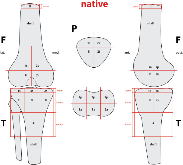
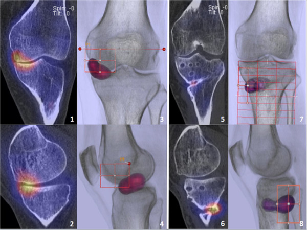
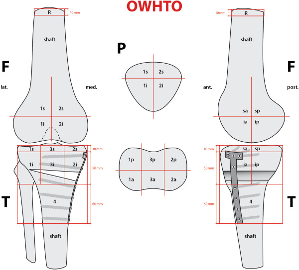
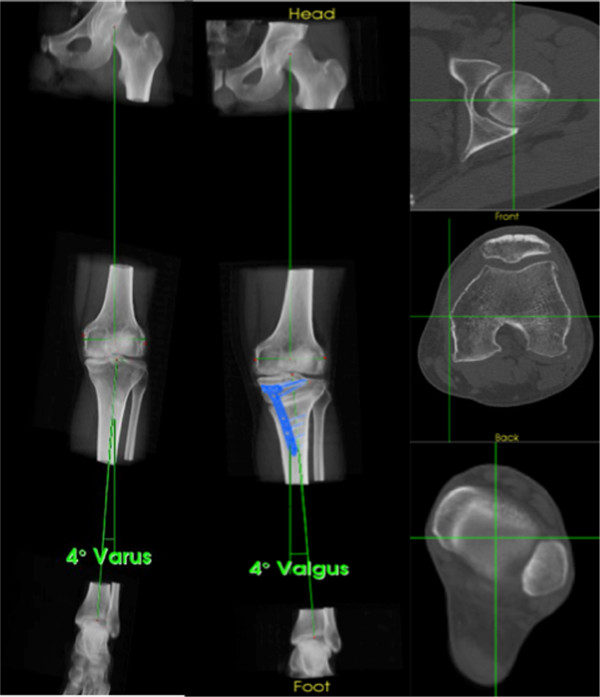
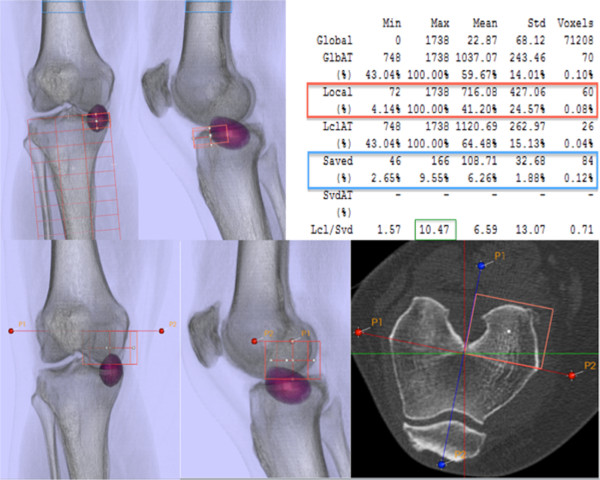
The localization scheme defines 9 femoral, 8 patellar, and 13 tibial zones to accurately map the examined tracer uptake volume in each anatomical area of interest. Maximum values for each area (mean ± standard deviation, median, and range) of the localization scheme were recorded as well as normalized values for the intensity of SPECT/CT tracer uptake calculated. The inter- and intra-OR was assessed for SPECT/CT localization and tracer activity. Pre- and postoperative mechanical alignment was assessed in SPECT/CT using a custom-made specialized software. The median inter- and intra-observer differences of the measured mechanical alignment were calculated along with the inter- and intra-OR.
The localization scheme showed near-perfect inter- and intra-OR (intra-class correlation coefficient (ICC) > 0.9) for the measurement of tracer activity and localization in all anatomical regions. For measurements of mechanical alignment, there was a strong agreement between the two observers (an inter-OR of ICC = 0.99 and an intra-OR of ICC = 0.98).
The presented SPECT/CT algorithm is highly reliable and clinically feasible. Combined with mechanical alignment analysis, it provides the surgeon with helpful information about realignment effects of high tibial osteotomies (HTOs) and might help identify the optimal personalized degree of correction in HTO surgery.
单光子发射计算机断层扫描和传统计算机断层扫描(SPECT/CT)示踪剂摄取已被证明反映了胫股膝关节的加载历史,并与机械和解剖学排列相关。我们的主要目的是为接受胫骨高位截骨术的患者开发一种新的标准化 SPECT/CT 算法,评估观察者间和观察者内的可靠性(OR),并评估其在胫骨高位截骨术前和术后患者随访中的临床适用性。
定位方案定义了 9 个股骨、8 个髌骨和 13 个胫骨区域,以准确地在每个感兴趣的解剖区域映射检查示踪剂摄取量。记录定位方案中每个区域(平均值±标准差、中位数和范围)的最大值以及计算的 SPECT/CT 示踪剂摄取强度的归一化值。评估 SPECT/CT 定位和示踪剂活性的观察者间和观察者内 OR。使用定制的专用软件在 SPECT/CT 中评估术前和术后的机械排列。计算测量的机械排列的观察者间和观察者内中位数差异以及观察者间和观察者内 OR。
定位方案在所有解剖区域中均显示出近乎完美的示踪剂活性和定位的观察者间和观察者内 OR(组内相关系数(ICC)>0.9)。对于机械排列的测量,两位观察者之间存在很强的一致性(观察者间 OR 的 ICC=0.99 和观察者内 OR 的 ICC=0.98)。
所提出的 SPECT/CT 算法具有高度可靠性和临床可行性。与机械排列分析相结合,它为外科医生提供了有关胫骨高位截骨术(HTO)重排效果的有用信息,并可能有助于确定 HTO 手术中最佳的个性化矫正程度。