Xing Xiaofang, Lian Shenyi, Hu Ying, Li Ziyu, Zhang Lianhai, Wen Xianzi, Du Hong, Jia Yongning, Zheng Zhixue, Meng Lin, Shou Chengchao, Ji Jiafu
Department of gastrointestinal translational research, Peking University Cancer Hospital & Institute, #52 Fu-Cheng Road, Hai-Dian District, Beijing 100142, China.
J Transl Med. 2013 Dec 13;11:309. doi: 10.1186/1479-5876-11-309.
PRL-3 is a member of phosphatases of regenerating liver family, characterized by phosphatase active domain and C-terminal prenylation motif. Overexpression of PRL-3 has been implicated in multiple cancers. Here we examined the clinical significance of PRL-3 in gastric cancer together with its metastatic biological functions utilizing different structural mutants.
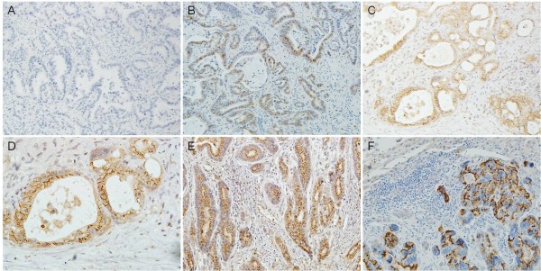
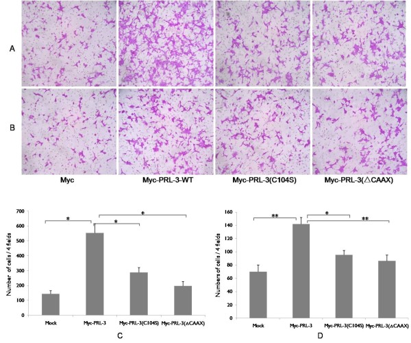
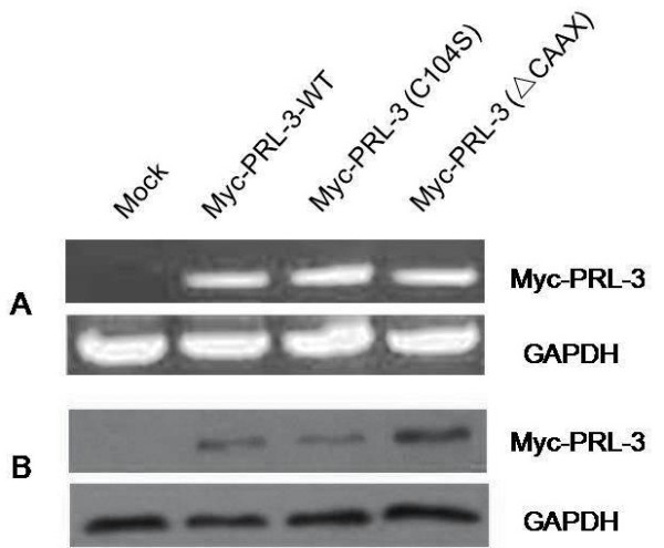
PRL-3 expression was analyzed immunohistochemically in 196 gastric cancer patients and 21 cases of liver metastasis. A series of wild type PRL-3 or its mutant plasmids were expressed in BGC823 cells to investigate the relationship between its catalytic activity, cellular localization and metastatic potential in vitro.
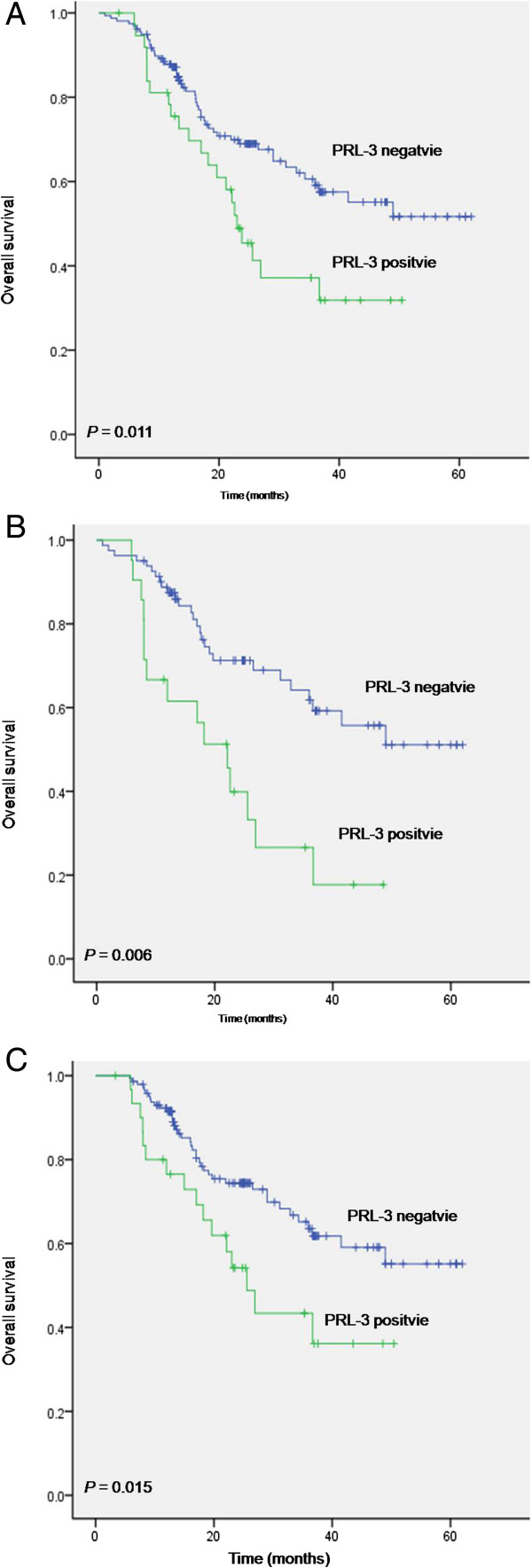
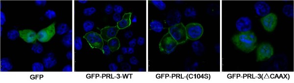
Positive staining of PRL-3 was observed in 19.4% (38/196) gastric cancer tissues compared with 76.2% (16/21) in liver metastasis. Statistical analysis revealed that PRL-3 expression correlated with lymph node metastasis and vascular invasion (P<0.05). Patients with high PRL-3 expression showed poorer 5-year overall survival (P=0.011). Wild type PRL-3 expressing cells resulted in enhanced migration and invasion ability, which were greatly crippled in form of PRL-3(C104S) or PRL-3(ΔCAAX) mutants accompanied with its alteration in subcellular localization.
Metastasis associated protein PRL-3 may serve as a potential prognostic biomarker in human gastric cancer. Both the phosphatase catalytic activity and cellular localization are critical for its function.
PRL-3是再生肝脏磷酸酶家族成员,具有磷酸酶活性结构域和C末端异戊二烯化基序。PRL-3的过表达与多种癌症相关。在此,我们利用不同的结构突变体研究了PRL-3在胃癌中的临床意义及其转移生物学功能。
对196例胃癌患者和21例肝转移患者的组织进行免疫组化分析PRL-3的表达。在BGC823细胞中表达一系列野生型PRL-3或其突变体质粒,以研究其催化活性、细胞定位与体外转移潜能之间的关系。
在196例胃癌组织中,PRL-3阳性染色率为19.4%(38/196),而在肝转移组织中为76.2%(16/21)。统计学分析显示,PRL-3表达与淋巴结转移和血管侵犯相关(P<0.05)。PRL-3高表达的患者5年总生存率较差(P=0.011)。表达野生型PRL-3的细胞迁移和侵袭能力增强,而PRL-3(C104S)或PRL-3(ΔCAAX)突变体形式则使其显著减弱,并伴有亚细胞定位改变。
转移相关蛋白PRL-3可能是人类胃癌潜在的预后生物标志物。磷酸酶催化活性和细胞定位对其功能都至关重要。