Ding Zhaohua, Newton Allen T, Xu Ran, Anderson Adam W, Morgan Victoria L, Gore John C
Vanderbilt University Institute of Imaging Science, Nashville, Tennessee, United States of America ; Department of Radiology and Radiological Sciences, Vanderbilt University, Nashville, Tennessee, United States of America ; Department of Biomedical Engineering, Vanderbilt University, Nashville, Tennessee, United States of America ; Department of Electrical Engineering and Computer Science, Vanderbilt University, Nashville, Tennessee, United States of America ; Chemical and Physical Biology Program, Vanderbilt University, Nashville, Tennessee, United States of America.
PLoS One. 2013 Dec 5;8(12):e82107. doi: 10.1371/journal.pone.0082107. eCollection 2013.
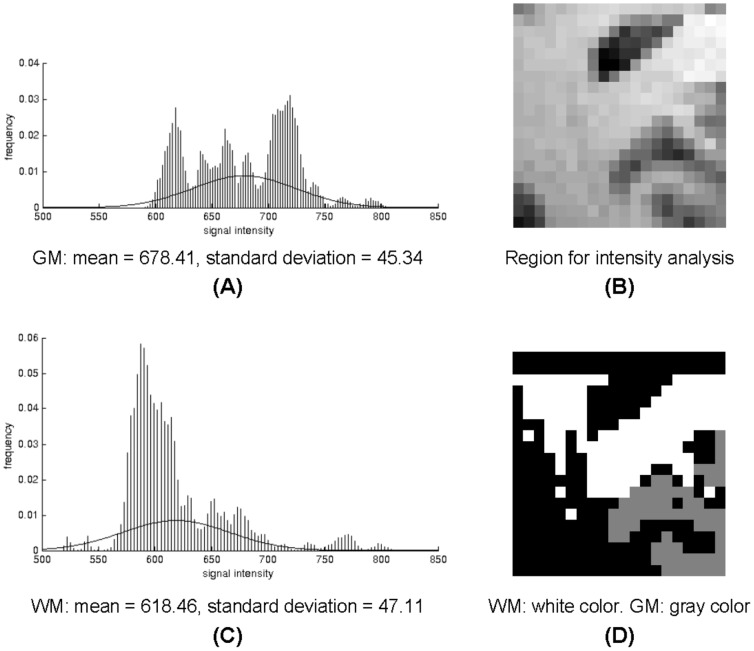
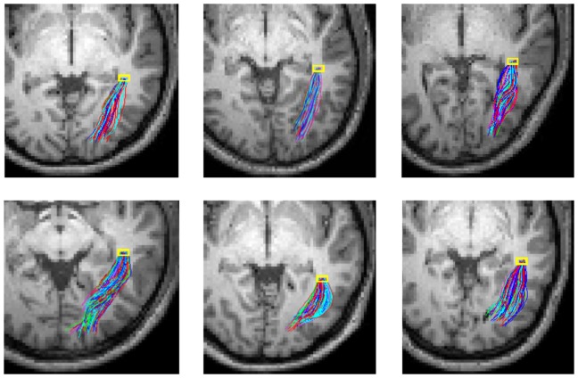
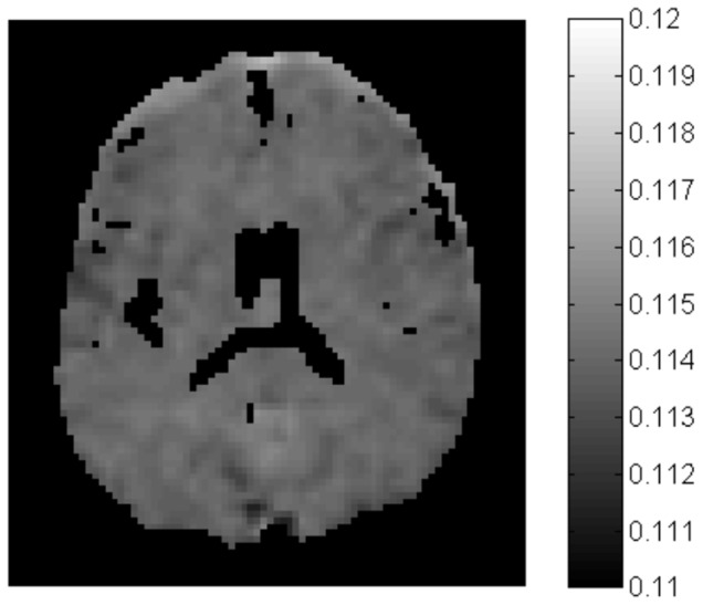
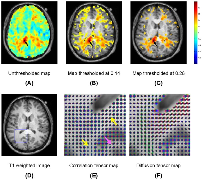
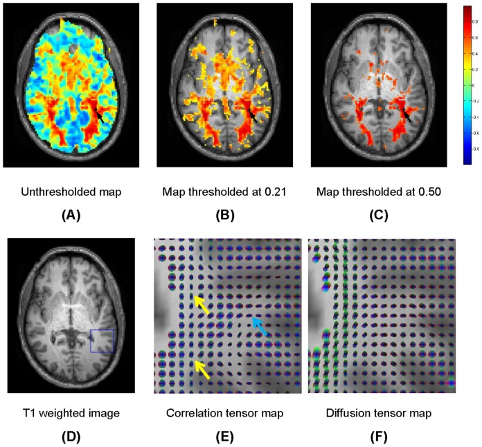
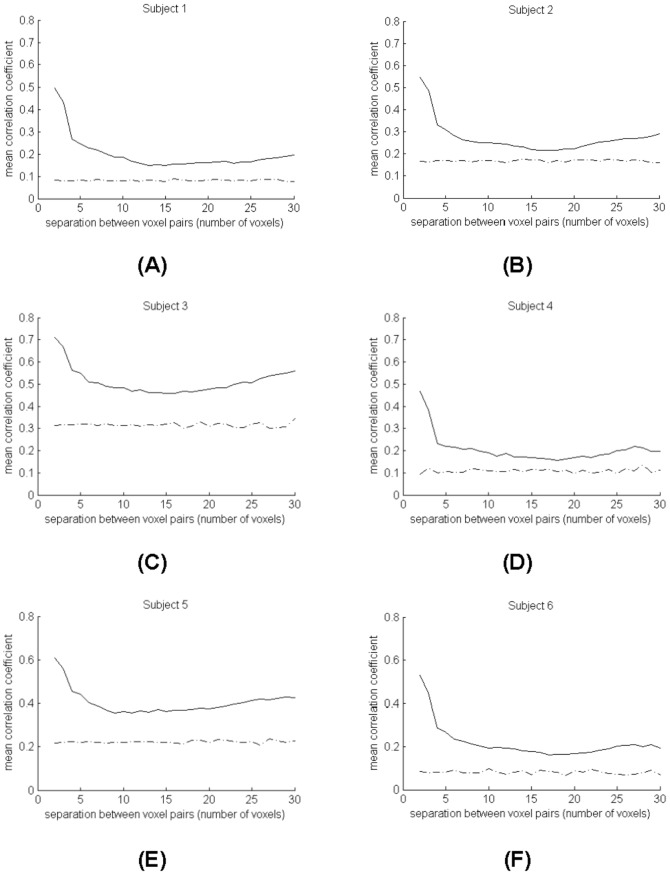
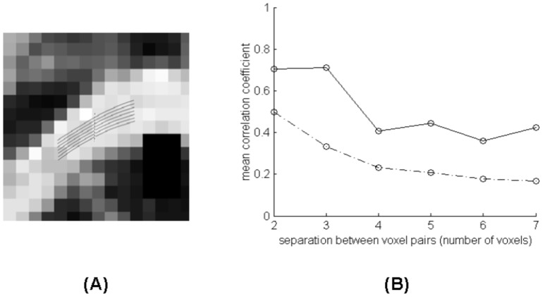
Resting state functional magnetic resonance imaging (fMRI) has been commonly used to measure functional connectivity between cortical regions, while diffusion tensor imaging (DTI) can be used to characterize structural connectivity of white matter tracts. In principle combining resting state fMRI and DTI data could allow characterization of structure-function relations of distributed neural networks. However, due to differences in the biophysical origins of their signals and in the tissues to which they apply, there has been no direct integration of these techniques to date. We demonstrate that MRI signal variations and power spectra in a resting state are largely comparable between gray matter and white matter, that there are temporal correlations of fMRI signals that persist over long distances within distinct white matter structures, and that neighboring intervoxel correlations of low frequency resting state signals showed distinct anisotropy in many regions. These observations suggest that MRI signal variations from within white matter in a resting state may convey similar information as their corresponding fluctuations of MRI signals in gray matter. We thus derive a local spatio-temporal correlation tensor which captures directional variations of resting-state correlations and which reveals distinct structures in both white and gray matter. This novel concept is illustrated with in vivo experiments in a resting state, which demonstrate the potential of the technique for mapping the functional structure of neural networks and for direct integration of structure-function relations in the human brain.
静息态功能磁共振成像(fMRI)已被广泛用于测量皮质区域之间的功能连接,而扩散张量成像(DTI)可用于表征白质束的结构连接。原则上,结合静息态fMRI和DTI数据可以表征分布式神经网络的结构-功能关系。然而,由于它们信号的生物物理起源以及所应用组织的差异,迄今为止这些技术尚未直接整合。我们证明,在静息状态下,灰质和白质之间的MRI信号变化和功率谱在很大程度上是可比的,在不同的白质结构内,fMRI信号存在长时间持续的时间相关性,并且低频静息态信号的相邻体素间相关性在许多区域表现出明显的各向异性。这些观察结果表明,静息状态下白质内的MRI信号变化可能与其在灰质中相应的MRI信号波动传达相似的信息。因此,我们推导了一个局部时空相关张量,它捕获静息态相关性的方向变化,并揭示了白质和灰质中的独特结构。这一新颖概念通过静息状态下的体内实验得到了说明,这些实验证明了该技术在绘制神经网络功能结构以及直接整合人类大脑结构-功能关系方面的潜力。