Monden Yu, Yamamoto Shohaku, Yamakawa Ryoji, Sunada Atsuko, Asari Seishi, Makimura Koichi, Inoue Yoshitsugu
Department of Ophthalmology, Kurume University School of Medicine, Fukuoka, Tokyo.
Laboratory for Clinical Investigation, Tokyo.
Clin Ophthalmol. 2013;7:2261-4. doi: 10.2147/OPTH.S48732. Epub 2013 Nov 27.
To report the isolation of Pestalotiopsis clavispora from the cornea of a patient with recurrent keratitis.
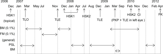
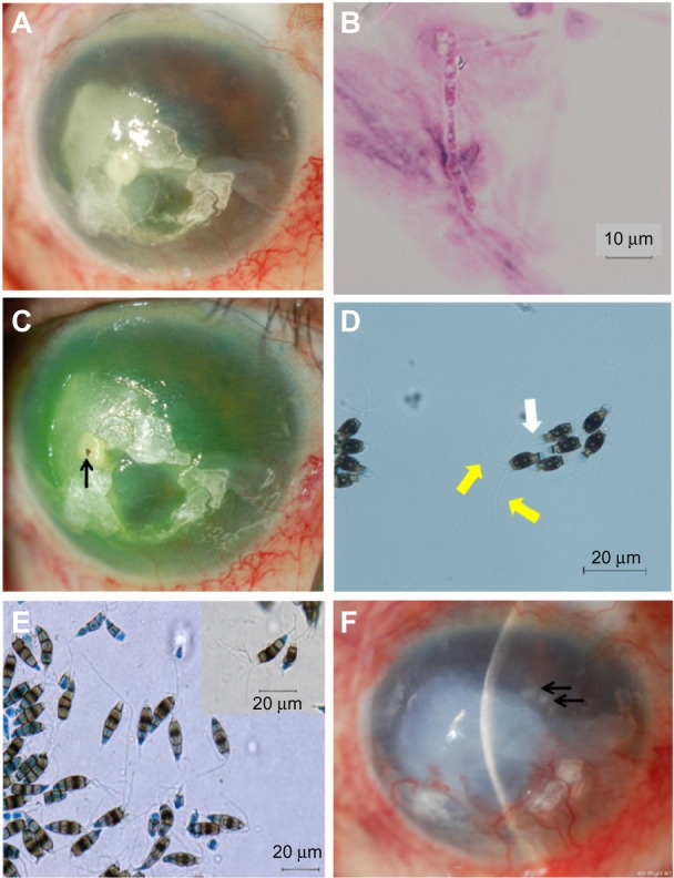
A 73-year-old male gardener presented with conjunctival injection and an oval infiltrate with feathery margins in the temporal half of the cornea in the right eye. His ocular history in the right eye included cataract surgery, five episodes of herpes simplex keratitis, three glaucoma surgeries, and bullous keratopathy. He had been treated with corticosteroids for years. Light microscopy of corneal scrapings revealed a filamentous fungus, and fungal keratitis was diagnosed. Treatment with topical voriconazole and pimaricin ointment was commenced. One month later, the infiltrate resolved. The antifungal agents were discontinued 7 months later, and keratitis relapsed 4 days after the discontinuation. The fungus was isolated and identified by molecular techniques as P. clavispora. Based on the results of antifungal susceptibility testing, treatment with topical and intravenous micafungin was initiated. The corneal infiltrate resolved 1 month after the relapse.
Molecular identification of the pathogen, and antifungal susceptibility testing, are useful in treating patients with fungal keratitis caused by a rare human pathogen.
报告从一名复发性角膜炎患者的角膜中分离出棒状拟盘多毛孢菌。
一名73岁男性园丁,右眼出现结膜充血,角膜颞侧有一个边缘呈羽毛状的椭圆形浸润灶。他右眼的眼部病史包括白内障手术、五次单纯疱疹性角膜炎发作、三次青光眼手术以及大泡性角膜病变。他多年来一直使用皮质类固醇治疗。角膜刮片的光学显微镜检查发现丝状真菌,诊断为真菌性角膜炎。开始使用局部伏立康唑和匹马霉素眼膏治疗。1个月后,浸润灶消退。7个月后停用抗真菌药物,停药4天后角膜炎复发。通过分子技术分离并鉴定出该真菌为棒状拟盘多毛孢菌。根据抗真菌药敏试验结果,开始使用局部和静脉注射米卡芬净治疗。复发1个月后角膜浸润灶消退。
病原体的分子鉴定和抗真菌药敏试验,有助于治疗由罕见人类病原体引起的真菌性角膜炎患者。