Williams Cassandra R, Dustin Michael L, Sauer John-Demian
Molecular Pathogenesis Program, The Helen L. and Martin S. Kimmel Center for Biology and Medicine at Skirball Institute of Biomolecular Medicine, New York, New York, United States of America.
Department of Medical Microbiology and Immunology, University of Wisconsin-Madison, Madison, Wisconsin, United States of America.
PLoS One. 2013 Dec 9;8(12):e83191. doi: 10.1371/journal.pone.0083191. eCollection 2013.
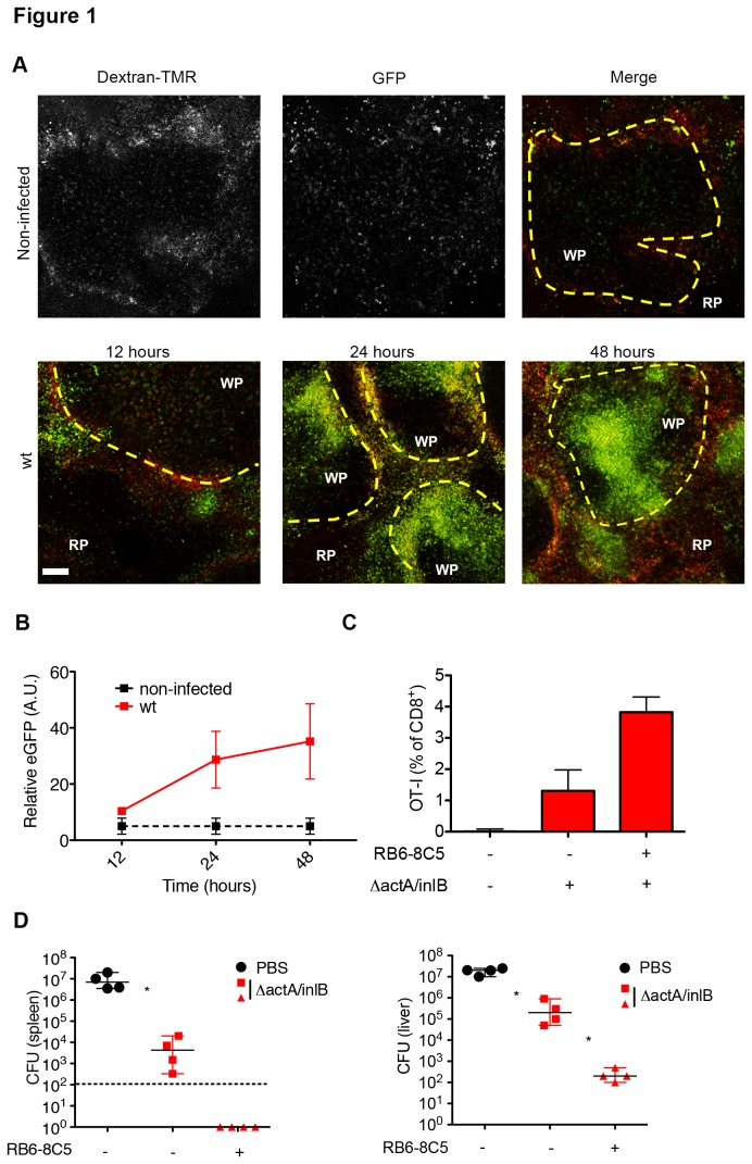
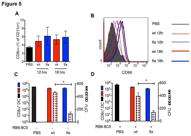
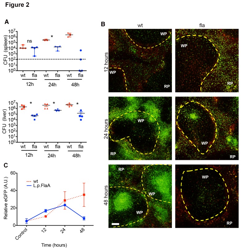
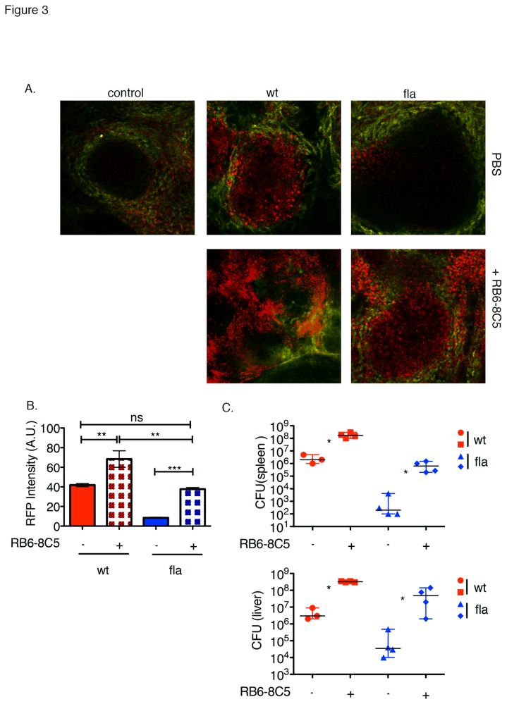
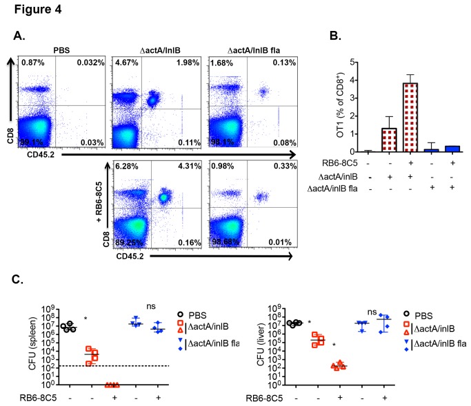
Activation of the Nlrc4 inflammasome results in the secretion of IL-1β and IL-18 through caspase-1 and induction of pyroptosis. L. monocytogenes engineered to activate Nlrc4 by expression of Legionella pneumophilia flagellin (L. monocytogenes L.p.FlaA) are less immunogenic for CD8(+) T cell responses than wt L. monocytogenes. It is also known that IL-1β orchestrates recruitment of myelomonocytic cells (MMC), which have been shown to interfere with T cell-dendritic cells (DC) interactions in splenic white pulp (WP), limiting T cell priming and protective immunity. We have further analyzed the role of MMCs in the immunogenicity of L. monocytogenes L.p.FlaA. We confirmed that MMCs infiltrate the WP between 24-48 hours in response to wt L. monocytogenes infection and that depletion of MMCs enhances CD8(+) T cell priming and protective memory. L. monocytogenes L.p.FlaA elicited accelerated recruitment of MMCs into the WP. While MMCs contribute to control of L. monocytogenes L.p.FlaA, MMC depletion did not increase immunogenicity of L.p.FlaA expressing strains. There was a significant decrease in L. monocytogenes L.p.FlaA in CD8α(+) DCs independent of MMCs. These findings suggest that limiting inflammasome activation is important for bacterial accumulation in CD8α(+) DCs, which are known to be critical for T cell response to L. monocytogenes.
Nlrc4炎性小体的激活通过半胱天冬酶-1导致白细胞介素-1β(IL-1β)和白细胞介素-18(IL-18)的分泌,并诱导细胞焦亡。通过表达嗜肺军团菌鞭毛蛋白(单核细胞增生李斯特菌L.p.FlaA)来激活Nlrc4的工程化单核细胞增生李斯特菌,相较于野生型单核细胞增生李斯特菌,对CD8(+) T细胞反应的免疫原性更低。还已知IL-1β协调骨髓单核细胞(MMC)的募集,骨髓单核细胞已被证明会干扰脾白髓(WP)中T细胞与树突状细胞(DC)的相互作用,从而限制T细胞启动和保护性免疫。我们进一步分析了MMC在单核细胞增生李斯特菌L.p.FlaA免疫原性中的作用。我们证实,响应野生型单核细胞增生李斯特菌感染,MMC在24至48小时内浸润WP,并且去除MMC可增强CD8(+) T细胞启动和保护性记忆。单核细胞增生李斯特菌L.p.FlaA引发MMC加速募集到WP中。虽然MMC有助于控制单核细胞增生李斯特菌L.p.FlaA,但去除MMC并未增加表达L.p.FlaA菌株的免疫原性。在不依赖MMC的情况下,CD8α(+) DC中的单核细胞增生李斯特菌L.p.FlaA显著减少。这些发现表明,限制炎性小体激活对于细菌在CD8α(+) DC中的积累很重要,而CD8α(+) DC已知对T细胞对单核细胞增生李斯特菌的反应至关重要。